Kirsi Syrlin

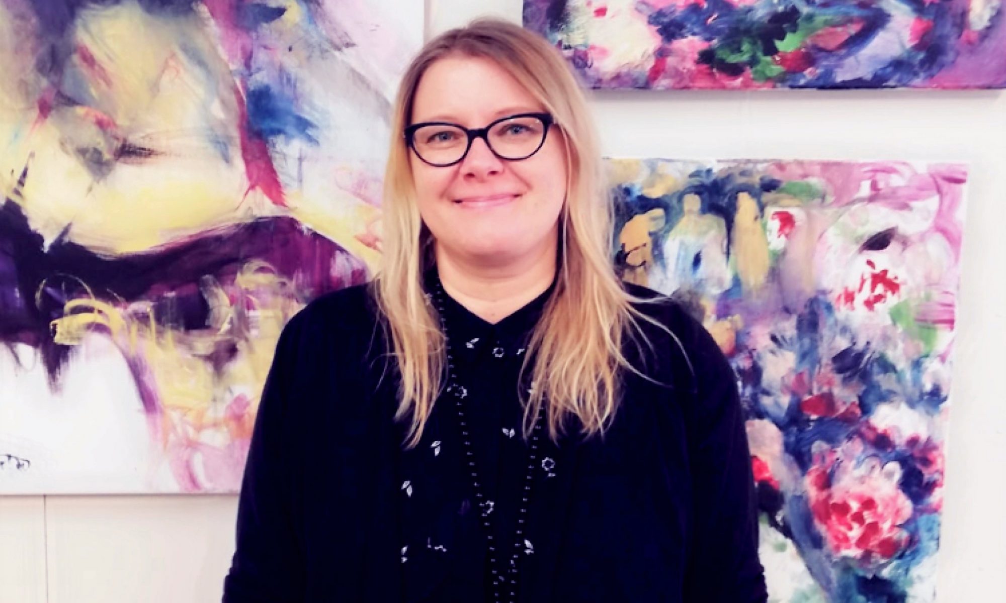
Kirsi Syrlin is a professional Finnish artist who has held exhibitions across Europe. 

Kirsi now resides in Espoo, Finland having returned from living in Belgium for several years. She has held exhibitions across Europe with more recent ones taking place in Finland, Italy, and Belgium. Her art can be seen in numerous private collections throughout Europe as well as closer to home in the City of Helsinki who have acquired her paintings for the Kivelä hospital. During her residency in Belgium, Kirsi held a solo exhibition at the Embassy of Finland in Brussels. Her time in Italy brought similar success which saw Kirsi’s pieces published in an Italian Artinside Magazine by curator Maddalena Grazzini. In addition, Kirsi was invited to participate in the Florence Biennale. Owing to this vast international exposure, Kirsi now has representation contacts with galleries in Italy as well as those further afield in the USA.

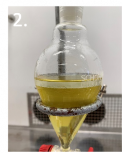
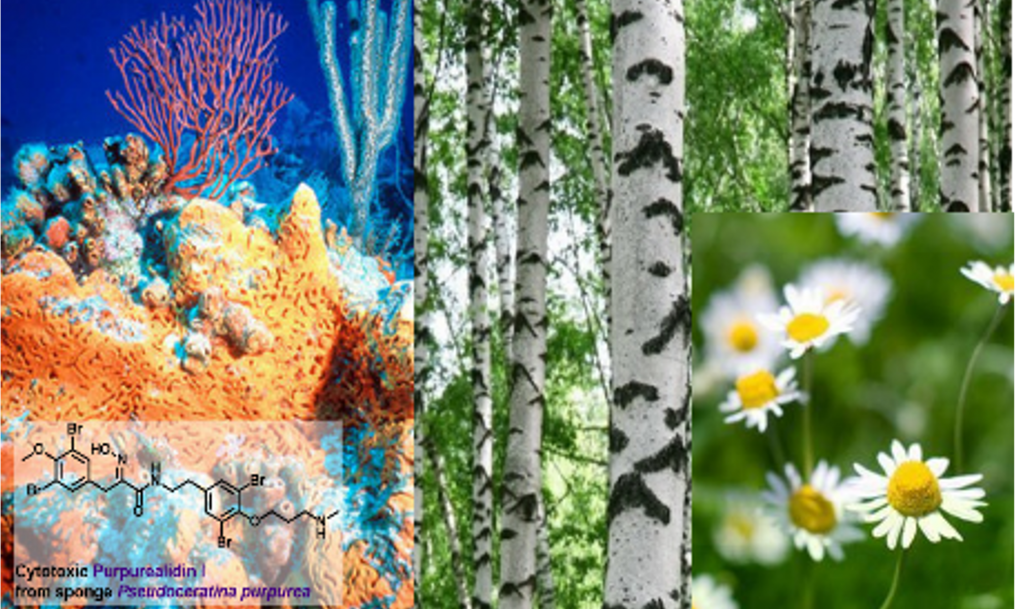
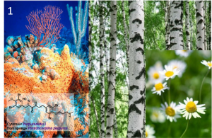
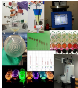
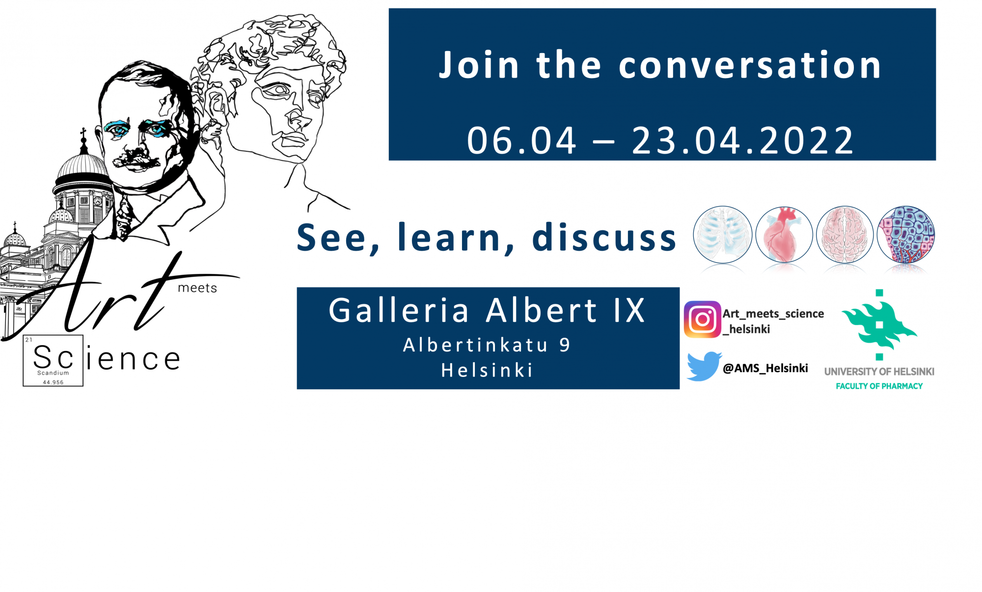
Reflecting on her reasoning for joining Art Meets Science, Kirsi shares her previous charity endeavours: “I have previously taken part in different charity auctions and projects and Art Meets Science sounded like a great opportunity to help the University of Helsinki’s Faculty of Pharmacy achieve their goal of sharing their research with the general public alongside raising funds for their professorship in sustainable pharmacy”. Kirsi continues, “the whole idea of combining art and science together is so inviting. They are so often compared as the opposite sides of the spectrum instead of powerful movements that could work best together”.

Looking ahead to the exhibition, Kirsi shares her excitement about seeing what the other collaborations have been working on together. Kirsi remarks “it is always so enchanting to see how the exhibition will look, how the light at the gallery moves around the pieces of art, and how the combination of different kinds of art pieces will create a unique moment for the opening evening. And of course, I am looking forward to meeting the other participants, the artists, scientists, & the audience in person”.

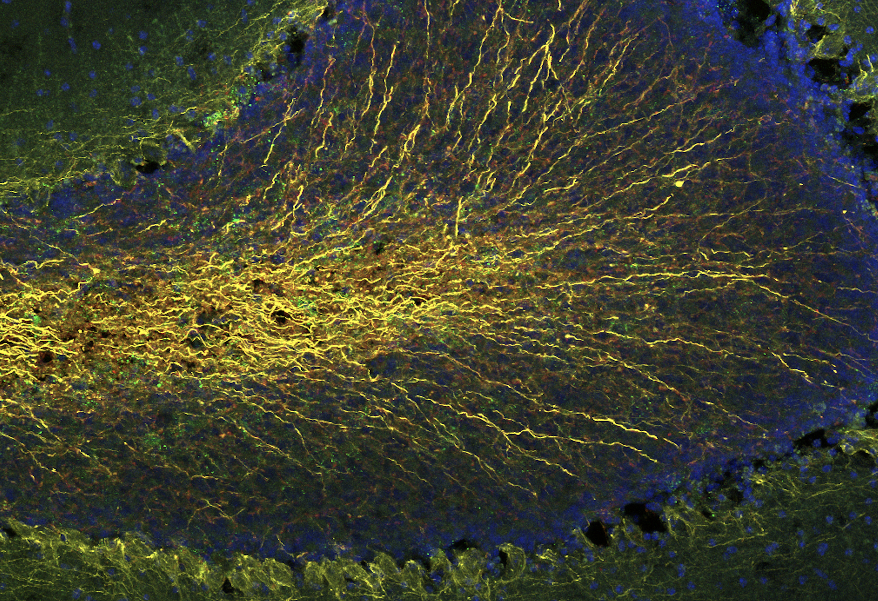
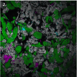
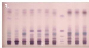
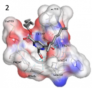
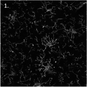
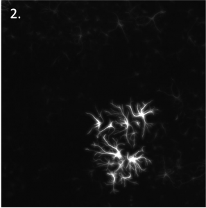
Kirsi has been collaborating with Dr Virpi Talman and Dr Qasim Majid of the Regenerative Cardiac Pharmacology group.

You can learn more about Kirsi and her work from her website and Instagram.

Regenerative Cardiac Pharmacology Lab

New Drugs for Heart Diseases

The human heart beats approximately 100,000 times every day. Pumping oxygen-rich blood across our body, the heart is essential in keeping our other organs healthy. A heart attack occurs when cardiac cells, including the beating heart muscle cells (cardiomyocytes) are themselves deprived of oxygen due to a blockage in a blood vessel in the heart. This in turn leads to the death of these cells which are incapable of regrowing, and therefore reduces the heart’s pumping capacity. This phenomenon is known as heart failure. Similarly, high blood pressure or genetic conditions can cause the heart muscle to stiffen or enlarge, decreasing the heart’s ability to pump blood.    

We aim to address some of the fundamental questions pertaining to heart diseases. One of our goals is to identify ways to force remaining heart muscle cells to regrow (proliferate) to heal the heart after a heart attack. Stem cells are excellent tools for such studies as they can become any cell type of the human body. In our lab, we generate heart muscle cells and blood vessel endothelial cells from stem cells and study the effects of different conditions, drugs, or chemical compounds on these cells.     

Learn more about Dr Qasim Majid and Dr Virpi Talman from the Regenerative Cardiac Pharmacology lab

Fluorescence microscope images of:

  1. Human pluripotent stem cell-derived cardiomyocytes.
  2. Human pluripotent stem cell-derived cardiomyocytes and endothelial cells.

Biopharmacy Research Group​

Prof. Yliperttula leads the multidisciplinary and multinational Biopharmaceutics research group at the University of Helsinki’s Faculty of Pharmacy.

The research program consists of the development of biomaterials for cell culture, drug delivery, and tissue repair together with detection technology. The developed biomaterials are based on nanofibrillated cellulose, nanoparticles, and extracellular vesicles. 

Two of the most important projects on-going in the Biopharmacy research group are:

  1. EV Ecosystem: https://www.bloodservice.fi/Research%20Projects/ev-ecosystem 
  2. GeneCellNano Flagship: https://www.genecellnano.fi 

Nanofibrillated cellulose in skin graft donor site treatment  (FibDex)

A.Detachment of dressing from donor site. without breaking the newly formed skin.

B.The epithelialised skin without scar formation.

In vitro and in vivo enhanced antitumor effects of oncolytic virus and paclitaxel encapsulated in extracellular vesicles (EVs).

A.Lung cancer cells were implanted subcutaneously into mice and EV formulations were administered intravenously (i.v.). Tumor growth was followed over time.

B.Kaplan-Meier test was used to calculate the survival profile.

Funding sources: BioCenter Finland, Tekes-The Finnish Funding Agency for Technology and Innovation, EU-FP7 (LIV-ES project, HEALTH-F5-2008-223317), Graduate School of Pharmaceutical Sciences, Finnish Red Cross Service, MATRENA graduate schools, Academy of Finland and EU-Erasmus Exchange Student Exchange Programme, UPM-GrowDexI I –III projects, UPM-Wound project, Huttunen foundation, professor-pool Orion foundation

Pharmaceutical Nanotechnology Lab

Light triggered drug release

A central theme in pharmaceutical research is controlling drug release. A particular area of interest for us is light-triggered release from nanosized drug carriers. We consider light to be the most flexible drug-release trigger for advanced drug delivery systems. Light can be used to both trigger and subsequently control the drug release with extreme time and location precision. To have the best tissue penetration, we are focusing on red-light activatable nanocarriers. Modern ultrathin light guides also enable the treatment of deeper tissues with minimal surgery.   ​​

A formidable physiological barrier to light-triggered drug release has been the inability to use high-energy blue/UV light as the triggering signal, making deeper targets within tissues accessible only to red light. However, red light, with its intrinsically lower energy, has limited value in photochemical reactions because e.g., the photocleavage of covalent bonds typically requires UV-light. In order to circumvent this issue, we focus on converting red light into blue light in precisely-tailored drug-releasing implants.   

Learn more about Prof. Timo Laaksonen and Dr Tatu  Lajunen from the Pharmaceutical Nanotechnology Lab.

Transporter group

After we take a drug, it will travel around the body in our blood, entering the cells of our different tissues, such as the liver. This movement can be helped, or hindered, by certain transport proteins. These transporters act like pumps on the cell membrane, protecting the cells from toxic compounds or bringing them essential nutrients. Their function can differ between individuals due to differences in genetics. Some drugs or dietary compounds may also alter their function. These changes can possibly affect the efficacy of drug treatment.

In our laboratory, we grow cells of human and animal origin and use them to study the function of transporters. When information about the function of a transporter is added to a computational model, we can predict its effect on drug treatment. In this way, we aim to ensure safe and effective drug treatments.

We have for instance studied how certain natural products and food additives can affect the function of transporters on a cellular level. Based on our results, we suspect that certain colourants, such as curcumin, may affect the absorption of drugs from the intestine. We have also studied the effect of genetic alterations on the function of several transporters. Many genetic alterations significantly decrease the function of transporters in cell studies, suggesting that drug levels in the body may be different in people with these alterations.

Learn more about Dr Eva Ramsay from Professor Heidi Kidron’s Transporter group.

  1. Graphic of a transport protein in the cell membrane, pumping drugs out of the cell and into the blood.
  2. Immunofluorescent microscope image of transporter proteins (green) on the surface of cells. The nucleus, where genetic information is stored, is in blue.

​Regenerative Neuroscience Lab

Regenerative Neuroscience: Developing  Regenerative Treatments for Incurable Diseases

The number of patients with neurodegenerative diseases such as amyotrophic lateral sclerosis (ALS), Parkinson’s disease (PD), and multiple sclerosis (MS) increases with age. There is no treatment that either prevents these diseases from progressing or repairs the damage to the nerve cells. ​​

The Regenerative Neuroscience lab is researching and developing new drug candidates for ALS, PD, and MS that prevent the disease from progressing and repair nerve cell damage. We use several different research models, such as stem cells, as well as various genetic and toxin-generated disease models. The goal of our research is to ascertain the most promising drug candidates and assess these in clinical trials for the benefit of patients.

Learn more about Tapani Koppinen and Aastha Singh from Professor Merja Voutilainen’s ​ Regenerative Neuroscience Lab.

Graphical illustration of:

  1. Amyotrophic lateral sclerosis
  2. Parkinson’s Disease
  3. Multiple sclerosis
  4. Modelling brain cell damage in brain slice cultures and searching for new therapies

ImmunoViroTherapy Lab

The Good Side of a Virus

Our research group develops vaccines against cancer. How do we do that? We modify viruses to seek and specifically destroy cancer cells whilst leaving live, healthy cells undamaged. During this process, we reprogram the immune system to store important information about the cancer to prevent it from emerging in the future. These special viruses are called oncolytic viruses and once combined with special features present in the tumour, they present a lethal strategy to destroy cancer. They work like a vaccine, teaching our bodies how to recognise emerging tumours as external intruders in advance of them becoming established, life-threatening cancers. 

Learn more about Manlio Fusciello from Professor Vincenzo Cerullo ImmunoViroTherapy lab.

Laboratory of Neurotherapeutics

The physiology underlying the effects of ketamine, psychedelics, laughing gas, and other rapid-acting antidepressants .

Depression is ever more common despite the growing investments in mental health care and the use of antidepressants. Conventional antidepressants alleviate the symptoms only after weeks of continuous use, and a third of patients do not benefit from them, although most suffer side effects. Thus, there is a large clinical demand for better treatments. However, to design new treatments, it is crucial to first understand how antidepressants work. 70 years since their discovery, there is still no verdict on how antidepressants alleviate the symptoms of depression.   

However, in contrast to conventional antidepressants, treatments like ketamine, psychedelics, nitrous oxide, electroconvulsive therapy, and sleep deprivation act within hours of a single treatment, with effects lasting up to weeks. As ketamine and laughing gas are removed from the body rapidly after administration, and electroconvulsive therapy and sleep deprivation are physiological in nature, it appears that the therapeutic response arises from the brain itself.  

Our research has uncovered a mechanism through which exposure to the brief treatment boosts brain plasticity and alleviates depressive symptoms. The mechanism is connected to the intrinsic regulation of sleep, energy, and metabolism. By understanding this phenomenon better, we may finally unravel how antidepressants work, opening new avenues for the development of more effective and better-tolerated treatments.  

Learn more about Okko Alitalo from the Laboratory of Neurotherapeutics.

  1. ​Summary of our research.
  2. Thermal camera portrait after a long day of experiments.
  3. Distribution of glucose in brains after laughing gas or medetomidine and thermal images of mice after laughing gas treatment.

Nanomedicines and Biomedical Engineering lab

Cross the cell membrane barrier: How smart polymers transfer therapeutics into cells  

All of the cells in our body are covered by protective membranes that prevent invasions from bacteria and viruses. The cell membrane does not allow big molecules, such as proteins or RNA, to enter freely into cells. 

Advances in modern medicine have seen the advent of advanced therapies such as gene therapy for the potential treatment of previously incurable genetic diseases. Further, mRNA vaccines against SARS-CoV-2, the virus responsible for the COVID-19 pandemic, have been developed as well as cell therapies for cancer treatment. All these treatments rely on the successful delivery of big therapeutic molecules into cells.  ​​

My research focuses on the development of smart polymer materials that function as delivery “vehicles”, that help therapeutic molecules cross the cell membrane barrier. I also use different techniques, trying to understand how these drug delivery “vehicles” interact with cells and transport within different compartments in the cell.   

Learn more about Dr Shiqi Wang and the Nanomedicines and Biomedical Engineering Lab.

  1. Illustration of how smart polymers transfer therapeutics across the cell membrane by “hijacking” the natural intracellular vesicles.
  2. Transmission electronic microscopy image of cell nuclei and natural intracellular vesicles.

Pharmaceutical Spectroscopy and Imaging group

Secrets of household items revealed by ​coherent Raman microscopy  

This project is a part of the Quantitative Chemically-Specific Imaging (qCSIInfrastructure for Material and Life Sciences, which offers highly advanced imaging technologies for industrial and academic use, in JyväskyläLappeenranta, and Helsinki campuses. In Helsinki, we have constructed a microscope unique to the Nordic countries. 

Unlike common light microscopy, our instrument uses so-called coherent Raman spectroscopy, which allows us to visualise the invisible infrared appearance of the sample. The infrared is a much broader and more information-rich spectral region than compared to the visible region we see with our eyes, therefore permitting chemically highly specific imaging. 

Learn more about Dr Teemu Tomberg from the Pharmaceutical Spectroscopy and Imaging group.

  1. Coherent Raman Microscope.
  2. Melatonin tablet as viewed under this microscope. Melatonin active ingredient, Magenta; bulking agents/stabilisers, green, cyan.
  3. Body Lotion as viewed under this microscope. Minor oily components form droplets in the surrounding water.

Bioactivity Screening Lab

In Search for Novel Antibacterials  

Bacteria are single-celled ancient creatures that are invisible to the human eye due to their small size. Nonetheless, millions of them surround us and inhabit our bodies in the same way that humans inhabit planet Earth. They are an integral part of human health, performing many important tasks such as aiding the digestion of food and processing nutrients. They also generate vitamins and form a protective layer on our skin.   

On the other hand, harmful bacteria can cause serious diseases. Antibiotic overuse over the last 80 years has resulted in the rapid emergence and spread of harmful, antibiotic-resistant bacteria, making common infections difficult, if not impossible, to treat. New bacteria-eliminating drugs and treatment methods are desperately needed.   

The Bioactivity Screening group at the University of Helsinki’s Faculty of Pharmacy employs high throughput screening to aid the discovery of antimicrobial drugs. We can test thousands of chemical compounds and natural products for their capacity to kill bacteria within days using multi-well plates and laboratory automation. In our lab, we also develop research tools. For example, the co-culture of bacterial and mammalian cells allows us to model the infection process and test novel ways to prevent infections.  

Learn more about Dr Polina Ilina from Professor Päivi Tammela’s bioactivity screening group.​

  1. Adding compounds to a 96 multi-well plate using a multi-channel pipette.
  2. Agar plates containing bacterial cells.

Bioactivity Screening Lab

Hidden Treasures: From Natural Products to Medicines

A natural product can be defined as a plant, an animal, or a microorganism, that has not been subjected to any treatment other than drying or other such preservation processes. A natural product can also be a part of an organism, for example, a leaf of a plant or an isolated organ of an animal.

Compounds derived from natural products can be obtained by extraction, followed by isolation and purification procedures. Isolated pure compounds are further tested for bioactivity by various methods. Many of the natural compounds are isolated, purified, and compounded directly into tablets or injectables. Drugs can also be based on derivatives of natural products where the chemical structure has been modified to obtain the desired medicinal properties. 

For centuries, natural products were the only available form of drugs. Among modern drugs, about 40% are of natural origin with some therapeutic areas seeing higher use of natural drugs. Approximately 60% of anticancer drugs and 75% of drugs against infectious diseases are either natural products or derivatives of natural products. 

At the University of Helsinki’s Faculty of Pharmacy, Prof. Tammela’s group studies various natural products for their chemical composition and bioactivity. One study area is the Chaga mushroom which is used in folk medicine for the treatment of cancer and gastric disorders. In the Bioactivity Screening lab, the Chaga mushroom is tested for its anti-herpes simplex virus ability.

Learn more about Dr Karmen Kapp from Professor Päivi Tammela’s bioactivity screening group.

  1. Chaga mushrooms in the forest.
  2. Extraction of natural products.
  3. Analysis of products.


Medicinal Chemistry Lab

The Medicinal Chemistry Lab designs, discovers, chemically synthesises, characterises, and optimises biologically active compounds.

Inspiration for the drug molecules can come from nature (plants, microorganisms, and marine compounds), computational molecular modelling, or repurposing previously known compounds.

Drug molecules are synthesised in the laboratory, purified, and then characterised. They are subsequently subjected to biological activity assays and the resulting biological activity information is used to optimise the compound further. We are synthesising small-molecule libraries of novel antibacterial, anticancer, antiviral, antiparkinsonian, and heart regenerating compounds. We are also developing green and sustainable methods to prepare the drug molecules.

Learn more about Dr Paula Kiuru from Prof. Jari Yli-Kauhaluoma’s Medicinal Chemistry lab.

  1. Identification of active compounds from nature.
  2. Computational design of new drugs.
  3. Synthesis of a new compound in the laboratory.


Neuroprotection and Neurorepair Lab

​Neuroprotection and Brain Repair

The brain is a complex structure that consists of various cell types like nerve cells (neurons) and supporting brain cells (glia). Our lab is interested in brain protection and repair processes thus; our research focuses on diseases like Parkinson’s disease and stroke. The central feature of neurodegeneration is a failure of proteins functioning correctly. Appropriate protein function is dependent upon the correct 3D protein structure. If this structure is disordered, the consequences for brain cells are often pathological. The problems in protein structure can act as a seed in the development of large aggregates and initiate disease progression, particularly in aged brains. Protein aggregates can be found in several neurodegenerative diseases and can be found within various compartments of a cell. We are also interested in the glia component of brain diseases. About half of the cells in the brain are cells other than the neurons. These are predominantly glial cells. A subset of these, the microglial cells, are small but mighty cells that allow the brain circuits to function correctly. They also mediate inflammation in the brain, which is a significant cause of pathology in many brain diseases. There are many aspects in glial cell biology that are still unknown, and it is likely future drugs will target these glial cells to treat neurodegenerative diseases. 

Learn more about Prof. Mikko Airavaara and Safak Er from Professor Mikko Airavaara’s Neuroprotection and Neurorepair lab.

Immunofluorescent microscopy images of:

    1. Microglia
    2. Astrocytes
    3. Dopamine neurone

Qasim Majid

Dr Qasim Majid is a postdoctoral researcher within the Talman lab and the initiator of Art Meets Science.

Originally from Manchester, Qasim has lived and studied in Newcastle and London. Having completed his PhD from Imperial College London, Qasim relocated to Helsinki at the height of the pandemic in 2020 to commence his postdoc.

His postdoctoral research work investigates the effect the cells of the blood vessels (endothelial cells) have on the beating heart cells (cardiomyocytes) and whether the former can cause the latter to grow and divide to replace the beating cells lost during a heart attack. Alongside his research activities, Qasim delivers lectures and supervises young researchers in the lab. Qasim remarks that “organising Art Meets Science on top of these many responsibilities for the last 1.5 years has therefore been challenging yet incredibly rewarding”.

When asked to share his inspiration behind starting Art Meets Science, Qasim discusses the misinformation campaign brought to the forefront during the COVID-19 pandemic: “misinformation continues to plague public discourse. As a man of science, I feel as though I am in a privileged position in which I can attempt to address this pandemic of mistruths. As such, I proposed Art Meets Science to my lab head, Dr Virpi Talman & thereafter the Dean of the Faculty, upon arriving at the University of Helsinki during the height of the COVID-19 pandemic in 2020”.

Reflecting on his experience thus far, Qasim remarks “It has been incredibly fun yet extremely challenging balancing the organisation of this event with my other duties. Nevertheless, it has been a pleasure corresponding with all the artists on a regular basis and learning about their work. Through this project, I have grown to understand pieces of art and the field of art a little better. To this end, one can draw similarities between the two (i.e., the creative process and the multiple iterations required to achieve the final form) as well as differences such as the pursuit of facts to completely understand something versus the ambiguity & personal interpretation afforded by art”.

Thinking ahead to the upcoming exhibition, Qasim is most excited to finally meet all the participants in person and engage with the public. Having invited school-aged children to the event, Qasim is hoping to inspire the next generation of scientists and artists. 

Qasim has been collaborating with Kirsi Syrlin and Gokhan Burhan.

You can read more about the research conducted within the Regenerative Cardiac Pharmacology group on their website.

Virpi Talman

Dr Virpi Talman is head of the Regenerative Cardiac Pharmacology group and a key player in the organisation of Art Meets Science:

Virpi describes herself as a “pharmacist by training and a researcher by spirit.”

As an Academy of Finland-funded Research Fellow in the division of Pharmacology and pharmacotherapy within the University of Helsinki’s Faculty of Pharmacy, Virpi leads the Regenerative Cardiac Pharmacology group who aim to discover new ways to treat heart diseases. In addition to her research activities, Virpi is also responsible for teaching undergraduate and postgraduate students in addition to supervising young researchers. 

Alongside Dr Qasim Majid, Virpi initiated Art Meets Science and has been a central light in the organisation of this event. This stemmed from a desire to promote discourse between science and the arts, both of which Virpi is highly passionate about, as well as wanting to find a way to discuss her research with the public in a way she hadn’t previously been involved in.

Reflecting on the project thus far, Virpi shares “organising the event has been a lot of work, but it has also been super inspiring! I loved meeting the artists with whom we collaborated, and I’ve been really encouraged by the positive feedback we have received”.

Looking ahead to the exhibition, Virpi shares her excitement for seeing the event come together and understanding how the artists have drawn inspiration from the research and channeled this into the pieces of art on display. Virpi continues and says “I am also looking forward to enjoying all the amazing art produced in the project and discussing our research with the public at the exhibition”.

Virpi has been collaborating with Kirsi Syrlin and Gokhan Burhan.

You can read more about the research conducted within the Regenerative Cardiac Pharmacology group on their website and via Virpi’s Twitter.

Gokhan Burhan

Gokhan Burhan is a Turkish-British artist residing in Finland. 

Gokhan is a visual artist and designer who creates collages, prints, and paintings with a focus on typography and abstraction. He has previously created typographic posters for election campaigns and charities.

When invited to participate in Art Meets Science, Gokhan was excited to use these techniques to showcase scientific research. Having experienced this first hand during this visit to the Talman lab, Gokhan enthused how “engaging and informative” an experience this was.

Gokhan has been collaborating with Dr Virpi Talman and Dr Qasim Majid of the Regenerative Cardiac Pharmacology group.

You can learn more about Gokhan and his work from his website and Instagram.

Marjo Yliperttula

Professor Marjo Yliperttula is a Professor of Biopharmaceutics at the University of Helsinki’s Faculty of Pharmacy. 

As a professor, Marjo is involved in teaching students throughout the academic ladder as well as leading her own research group.

When asked for her rationale behind participating in Art Meets Science, Marjo shares “I found this project to be very inspiring and a great way to share the research we conduct at the University with the public. Thus far, the project has been extremely interesting and stimulating, and has allowed me to get to know a range of new people including researchers and artists”.

Looking ahead to the exhibition, Marjo is hoping to see a great number of visitors at the exhibition with open minds and questions”.

Marjo has been collaborating with Petra Kaminen Mosher and Hannele Rekola.

Hannele Rekola

Hannele Rekola is a laboratory nurse and artist. 

Hannele is originally from Rauma but now lives in Järvenpää where she works at Meilahti Pathology as a laboratory nurse. In her free time, Hannele practices pottery and draws a Kwaakku cartoon that is published in Kaupunkilehti Raumalainen. Owing to her artistic talents, Hannele belongs to the Järvenpää Art Society.

Reflecting on her participation in Art Meets Science, Hannele remarks how she was inspired by her good friend, Professor Marjo Yliperttula, a professor at the University of Helsinki’s Faculty of Pharmacy, who informed Hannele of the project. Hannele continues “Marjo invited me to participate in the project and I immediately grew excited. Ideas started spinning in my head! My original idea of ​​ceramic vesicles was born and has since been transformed into a piece of art called ‘Connecting’. In this piece, vesicles play an important role in the pulsating umbilical cord that unites women”.

When asked about her experience thus far, Hannele shares how inspiring and necessary such a project is: “this joint project of science and art opens up huge new avenues to bring out the achievements of science through the eyes of art. It creates a third dimension that serves as a new kind of bridge for the viewer and an entry into the world of science that can sometimes be out of reach for the layman. Likewise, it acts as a reverse gateway to art, the opportunity to see things differently, through the eyes of art”.

Thinking ahead to the exhibition, Hannele is excited to see the reactions of people and the public alike towards this innovative and groundbreaking exhibition that combines science and art. Hannele remarks, “I very much hope that we will gain visibility and reach the audience and the media. In addition, I hope for interesting encounters with people, both those involved in the project and those coming to see the exhibition”.

You can learn more about Hannele and her work from her website and Instagram.

Petra Kaminen Mosher

Petra Kaminen Mosher is a Finnish-American artist living and working in Oulu, Finland.   

Originally from New York City, Petra received her art degree from Boston University prior to moving to Finland where she works as a full-time visual and textile artist, and portrait painter.   

When asked to specify what inspired her to join Art Meets Science, Petra shared how she has “always had a general interest in science and scientific development, particularly in medical innovation. Various scientific fields have inspired my work throughout my career, such as biology, botany, and pathology. I feel that the appreciation and understanding of scientific endeavours should be fostered and encouraged in the general public, and science collaborating with the arts is a stimulating and productive way to achieve that goal”.  

Petra has been collaborating with her long-term friend,  Prof. Marjo Yliperttula, Professor of Biopharmaceutics at the University of Helsinki’s Faculty of Pharmacy. Reflecting on this experience, Petra says “ It has been a continuous rumination on the interconnectedness of the natural world and the evolution of medicine.” 

Thinking ahead to the exhibition, Petra is looking forward to experiencing how the scientific impact of medical innovation is rendered into emotive visual media. 

You can learn more about Petra and her work from her website and Instagram.