Discovering the potential of next-generation probiotics (NGPs) as a HiLIFE trainee


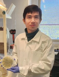
I’m Pinja Perkkiö, an aspiring microbiologist and a HiLIFE scholarship trainee in Reetta Satokari’s lab (University of Helsinki, Faculty of Medicine)! I’ve just finished my bachelor’s degree and I will start my studies in the master’s programme of Microbiology and Microbial biotechnology next autumn. This summer, I will be diving deep into the world of gut microbiota as I research the microbiota restoring potential of NGPs in intestinal inflammation and dysbiosis. 


How antibiotics disturb our gut microbiota 

The gut microbiota is defined as a community of microbes, e.g. bacteria, fungi, viruses and archaea residing within the gut. The microbiota is responsible for many important functions, such as breaking down complex carbohydrates and protecting the gut from infections. When a person takes antibiotics, it not only kills the bacteria causing the disease, but also the beneficial ones as antibiotics aren’t strain-specific. This allows potentially pathogenic bacteria to grow in larger numbers – the situation is called dysbiosis, meaning that the microbial community composition has been altered in a harmful way. 

Luckily, we have a tight layer of cells – the epithelium – that lines the walls of the gut and is covered by two layers of mucus. The mucus combined with the epithelium acts as a barrier between our intestines and the rest of our body, preventing intestinal pathogens from causing too much harm. Normally, it is very difficult for microbes to cross the mucus layer as it traps microbes and is loaded with antibacterial substances. In addition, the cells of the epithelium are tightly attached to each other, preventing any further movement of microbes. There are also lots of immune cells embedded in gut tissues that are ready for action, should a pathogen try to cause an infection. 

However, as dysbiosis leads to the growth of pathogenic bacteria, our immune cells (neutrophils, NK cells and macrophages, for example) activate inflammatory reactions. While these reactions are necessary for the elimination of pathogens, they also damage gut tissue. Pathogenic bacteria also activate the secretion of inflammatory molecules, cytokines, in a type of gut epithelial cell called enterocyte, which leads to the epithelium getting “leaky” – this means that microbes can cross the epithelium more easily and cause further inflammation in gut tissue. These bacteria can not only cause nasty infections, but they may also travel deeper in gut tissue or even to other sites in the body, increasing the risk of certain cancers (Bastos et al., 2023; Kouzu et al., 2022)! 


Next-generation probiotics against dysbiosis 

To fight antibiotic-induced dysbiosis and reduce the inflammation and leaky gut syndrome caused by it, researchers have turned their attention to potential new bacterial strains that could function as next-generation probiotics. They are defined as “live microorganisms identified on the basis of comparative microbiota analyses that, when administered in adequate amounts, confer a health benefit on the host” (Martín R, Langella P., 2019) – in simpler terms, microbes that can potentially help boost our health. Traditional probiotics, such as Bifidobacteria and Lactobacilli, have shown great potential in restoring a healthy microbial community in the gut after taking antibiotics, thus preventing dysbiosis and infections. They have also been shown to inhibit the production of inflammatory molecules in both immune cells and epithelium and boost the production of anti-inflammatory molecules.  


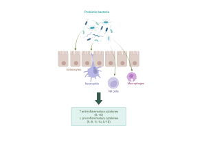
Probiotics exert anti-inflammatory effects on several cell types. Created with Biorender.com. 


There’s still lots of research to be done to bring next-generation probiotics into wide use. More information is needed on the different strains of potential probiotic bacteria and their specific effects on both the immune system and the gut epithelium. This is where my research project comes in! 

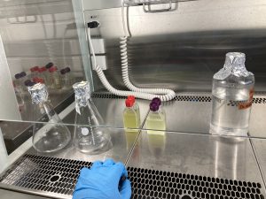
The project I’m collaborating on focuses on a mix of 14 different anaerobic bacterial strains that have the potential to restore the gut microbiota after antibiotic use and prevent or reduce inflammation. The next-generation probiotic mix was originally tested in an in vitro mucosal-simulator of the human gastrointestinal tract (M-SHIME(R)) model. A natural gut microbiota was obtained from the stool sample of a healthy human donor, after which it could grow. Then, the “gut” was exposed to antibiotics to induce dysbiosis. After the antibiotic treatment, one system was treated with the probiotic mixture and the other one with a negative control. Finally, samples from probiotic- and control-treated microbiotas were taken. Bacteria present on these will be studied to check the impact of the antibiotic intake in the microbiota and compare its possible recovery after the probiotic treatment. The production of short-chained and branched fatty acids, lactate and ammonia at different time points will also be measured since their levels indicate how much beneficial bacteria there are compared to harmful ones. 

To study how the anaerobic isolates might affect intestinal inflammation, I will culture two different intestinal epithelial cell lines: Caco-2 and HT-29. These cell lines were chosen because they are well-characterized and resemble enterocytes on the gut epithelium. Then, I will expose the HT-29 cell line to either the anaerobic bacterial blend or a placebo. After incubation, pro-inflammatory IL-8 production in the HT-29 cell line will be measured using ELISA assay. Later, the capability of a single freeze-dried strain in attenuating the IL-8 levels produced by HT-29 cell line will be assessed.  

Caco-2 cell line will be used to measure the effect of the microbiota samples collected from M-SHIME(R) on the barrier integrity of the Caco-2 (enterocyte) monolayer. The integrity will be assessed by measuring the TEER (transepithelial/transendothelial electrical resistance) of the cell monolayer, and the results will tell us if our strains can alleviate leakiness in the epithelial cell layer caused by dysbiosis. 


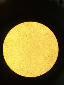
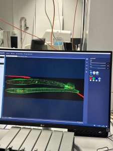
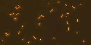
HT-29 cells under a microscope. 


Starting my research 

So far, I’ve read lots of literature related to the gut microbiota and treating inflammation with probiotics and next-generation probiotics. This has given me a good foundation on which to build my knowledge. The steps using M-SHIME(R) human gut simulator have been completed previously, so I’ve mainly been growing the intestinal cell lines required for our experiments. Luckily, I already have some experience with cell culture, so the start hasn’t been too difficult. My group has also been supportive and helpful, so I’m optimistic about the upcoming months! 


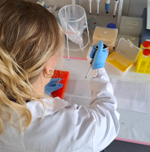
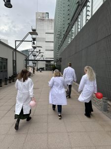
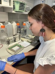
Seeding HT-29 cells for our experiments. 


The next step will be starting the IL-8 induction experiments, and once the initial results are ready, we can proceed to plan further. If you’re interested in our results, stay tuned for my next blog post! 


Sources: 

Bastos AR, Pereira-Marques J, Ferreira RM, Figueiredo C. Harnessing the Microbiome to Reduce Pancreatic Cancer Burden. Cancers (Basel). 2023 May 5;15(9):2629. doi: 10.3390/cancers15092629. PMID: 37174095; PMCID: PMC10177253. 

Kouzu K, Tsujimoto H, Kishi Y, Ueno H, Shinomiya N. Bacterial Translocation in Gastrointestinal Cancers and Cancer Treatment. Biomedicines. 2022 Feb 4;10(2):380. doi: 10.3390/biomedicines10020380. PMID: 35203589; PMCID: PMC8962358. 

Martín R, Langella P. Emerging Health Concepts in the Probiotics Field: Streamlining the Definitions. Front Microbiol. 2019 May 21;10:1047. doi: 10.3389/fmicb.2019.01047. PMID: 31164874; PMCID: PMC6536656. 


Some literature: 

Berta Bosch, Saliha Moutaharrik, Andrea Gazzaniga, Kaisa Hiippala, Hélder A. Santos, Alessandra Maroni, Reetta Satokari. Development of a time-dependent oral colon delivery system of anaerobic Odoribacter splanchnicus for bacteriotherapy. European Journal of Pharmaceutics and Biopharmaceutics. 2023; 190. 73-80. https://doi.org/10.1016/j.ejpb.2023.07.010. 

Cristofori F, Dargenio VN, Dargenio C, Miniello VL, Barone M, Francavilla R. Anti-Inflammatory and Immunomodulatory Effects of Probiotics in Gut Inflammation: A Door to the Body. Front Immunol. 2021; 26(12):578386. doi: 10.3389/fimmu.2021.578386.  

Marzorati M, Van den Abbeele P, Bubeck SS, Bayne T, Krishnan K, Young A, Mehta D, DeSouza A. Bacillus subtilis HU58 and Bacillus coagulans SC208 Probiotics Reduced the Effects of Antibiotic-Induced Gut Microbiome Dysbiosis in an M-SHIME® Model. Microorganisms. 2020; 8(7):1028. https://doi.org/10.3390/microorganisms8071028 

Succesfull lauch of HiLIFE’s Reseach Intensive Study Module

Group photo with the Research Intensive Study Module students & organizer Juha Saarikangas.

The first HiLIFE research training module was recently completed and proved to be an immidiate success. Organized in collaboration with the Genetics and Molecular Biosciences Master’s program (GMB), this module is designed to equip master’s students for future PhD studies. Comprised of lectures (GMB-901) and hands-on research (GMB-902) the module offered a blend of knowledge and experiences for students who are interested in continuing on the academic path.

Ten exceptional students, Eli, Georgios, Nea, Neža, Nicholas, Nisa, Nuša, Vandhana, Veikka and Vid were chosen based on their academic merits and compelling motivation letters to embarked on an research journey. They were each partnered with a HiLIFE-affiliated research groups or technology centers in one of the HiLIFE’s operative units — BI, FIMM, or NC — across three campuses where the students delved into diverse research projects. The spectrum of research was broad, ranging from establishing CRISPR knockout libraries for studying colorectal cancer development, to exploring the effects of small molecules on pancreatic beta cells, and molecular simulation studies aiming to unravel the function of the respiratory chain of mitochondria.

Nicholas presenting his project.
Nea carrying out her research work.

Once a week, we would gather for ‘group meetings,’ during which students presented their projects and the techniques they had learned in their respective host groups. These sessions facilitated knowledge exchange, presentation-, and problem-solving skills, overall fostering a collaborative camaraderie among the students.

The course culminated in a poster session on Fri 10.5.2024 at the Viikki Campus, where students presented their findings to an audience comprising fellow students and more senior researchers. The poster evaluation committee, comprised of GMB director Kari Keinänen and BI director Juha Huiskonen, lauded the overall quality of research conducted during the internships. The best poster price was awarded to Eli Eriksson from the Battersby lab for her presentation titled “Co-translational errors in mitochondrial protein synthesis are linked to cell fitness.” As a reward, Eli secured a prestigious HiLIFE Research Trainee Scholarship including 4-month salary to support her master’s thesis research.

Heartfelt congratulations to Eli and all participating students for their remarkable research achievements within a constrained timeframe. Also, big thanks to all the participating research groups who provided fascinating projects and excellent guidance to the students.

The call for research groups and students to partake in the 2025 course is set to open in late fall. Don’t miss out participating in this fantasticly rewarding journey!

Juha Saarikangas, Organiser

Eli Eriksson and the winning poster.
Poster session at Biocenter 2, Viikki Campus.

Beneath the surface: Unveiling the hidden dynamics of a Baltic ringed seal population


I’m Murat, a second year student in the Master’s programme in Life Science Informatics at the University of Helsinki, specializing in Biomathematics and Biostatistics. Last summer I embarked on a 4-month internship with the Environmental and Ecological Statistics group that was funded by HiLIFE, where I was able to apply my quantitative skills to a field I have been passionate about since I was a child: wildlife conservation. And what a journey it has been! Although my internship officially ended in September, I have kept working on the project until now.

My mission was to develop a Bayesian state-space model to help monitor Baltic ringed seals, a sub-species of ringed seals that was once on the verge of extinction due to unsustainable hunting practices and large-scale reproductive failure caused by pollution. Thankfully, the population has been recovering during the last few decades, at least in the northern region of the Baltic Sea¹. However, highly dynamic conditions brought on by changing sea ice patterns, decreasing pollution levels and the recent re-introduction of seal hunting have made reliable population monitoring increasingly difficult, and assessments of population status have not been possible for over a decade. The inability to estimate population size and growth rate has been a major obstacle in determining sustainable management practices.

During the summer and fall, ringed seals spend most of their time feeding out at sea, trying to accumulate fat reserves for the winter (foraging period). It is common for young seals to accidentally become entangled in fishing gear during this time. As the Baltic sea begins to freeze over, seals construct snow lairs on the sea ice for protection from harsh weather and predators (subnivean period). Adult females give birth to a single pup inside the snow lair. Once the snow and ice begin to melt, seals move out of their lairs and spend most of their time molting on the ice under the sun (molting period). This is the only time when ringed seals are easily visible, and aerial surveys are conducted each year in the spring to count the number of seals basking on ice. [2] (The illustration was created with the aid of DALL-E 3.)
My challenge was to create and parametrize a model that could accommodate all of these changing factors as well as the uncertainties associated with them, something conventional monitoring methods fell short of. Instead of analyzing different data sources separately, I integrated them into a single, unified model of ringed seal population dynamics – a method that is often called integrated population modeling³. By analyzing all available data holistically, integrated population models can exploit synergies between different data sources, making it possible to parametrize detailed and mechanistic population models.

The results we obtained with this approach were as exciting as they were important. For example, we discovered that the reproductive rates of seals might have fully recovered from the effects of past pollution, and the population may have increased from less than 5,000 to nearly 30,000 individuals! We also discovered that the recent re-introduction of seal hunting has had a notable impact on population growth, though not critical enough to ring any alarm bells for the seal population—yet. Another interesting finding was that seals may be behaving differently during years with low sea ice cover, hauling-out on ice in larger numbers during their annual molt in the spring. Since hauled-out ringed seals are counted each year during this time, population counts after warm winters may be significantly higher! This means that as sea ice patterns are altered due to climate change, reliably estimating the population size could become increasingly more difficult.

A photo of my research group on a beautiful summer day, after an all-day boat party with swimming, sauna and other fun activities.

During the time I was working on this project, every day brought new and diverse challenges – a rich blend of mathematics, statistics, and ecology. Collaboration with researchers from other fields was not just helpful, but essential, making my days anything but repetitive. A year of hard work has finally culminated in a manuscript that is ready to be submitted for publication at a scientific journal! Reflecting back at the journey, it is amazing to see how much I have learned. I started my internship with many doubts – unsure whether I would enjoy doing research, and whether I had what it takes. I am now seriously considering a PhD, confident that a career in research is right for me. As I near the end of this project, I am filled with gratitude for the opportunity to have played a role in preserving these amazing animals, and I eagerly anticipate my next endeavor in the world of research.

References:

¹Sundqvist, L., Harkonen, T., Svensson, C. J., and Harding, K. C. (2012). Linking climate trends to population dynamics in the Baltic ringed seal: Impacts of historical and future winter temperatures. Ambio, 41:865–872.

²Kelly, B. P., Bengtson, J. L., Boveng, P. L., Cameron, M. F., Dahle, S. P., Jansen, J. K., Logerwell E.A., Overland J.E., Sabine C.L., Waring G.T., & Wilder, J. M. (2010). Status review of the ringed seal (Phoca hispida).

³Schaub, M. and Abadi, F. (2011). Integrated population models: a novel analysis framework for deeper insights into population dynamics. Journal of Ornithology, 152:227–237.

How my experience as a HiLIFE trainee brought out the bookworm inside me

My HiLIFE traineeship has come to an end, and although it is sad to close a chapter, I couldn’t be more excited to share how my experience went.

I am Mireia Pagès Guitart, I just graduated with an MSc in Pharmaceutical Research, Development, and Safety at the University of Helsinki. In September 2023, I started an incredible project at Michael Jeltsch’s lab to tackle a huge sustainability challenge with one of the best tools at our disposal: science. You can learn more about it by reading my previous post, by clicking here. In January 2024, HiLIFE supported my investigation by awarding me the HiLIFE research traineeship. Do you wonder how it ended up? Keep reading!

After months of reading, testing, failing, achieving and, most importantly, having fun, I am happy to share that I succeeded in expressing GFP in vitro. Unexpectedly, it turned out that earthworms have inherent green fluorescence. Therefore we could not be sure whether the green color after in vivo imaging was due to our protein or due to the animal. Even so, my project was a big step forward to achieve our goal: developing a sustainable animal model for protein production and microplastic degradation. You might wonder: what now? I will continue my journey in Catalonia (for now) and my lab team will continue this amazing project… so be aware of a promising future!

Worm Imaging. How cool is it?

The HiLIFE traineeship made me realize I enjoy research more than I thought! Doing extra hours, having to check up on the cells during weekends, and messing up experiments doesn’t matter if you have science at your heart. During my experience, I thought about the thin line between a job and a hobby, and how my project became my pastime. Feeling this way towards research couldn’t have been possible without being surrounded by a comforting team. I am aware of how lucky I was to find a lab that matched my energy, sense of humor and enthusiasm for research.

Persian dinner by my colleague Honey, with my lab group. So delicious…

Joining a research group goes beyond the lab work. I was proud to join workshops, competitions, conferences and talks that were as important to grow as a scientist. The value of grasping every opportunity during my stay brought out the bookworm inside me. Nothing but good outcomes in my research and personal progress came afterwards. I encourage future HiLIFE trainees to make the most out of their traineeship.

Our lab team as “The Bunsen Burners” for the Research Program Unit competition 🙂

Over my master’s at the University of Helsinki and my stay at the lab, I have internalized many learning lessons from which I want to share three:

  1. Whatever you do, stay curious and skeptical, and, above all, enjoy the journey because there is no other destination (by my PI Michael).
  2. Sometimes you think you have been buried, but actually, you have been planted (by the activist Christine Caine)
  3. Surround yourself with people who are different from you: open your eyes and envision your view of this small world with big souls (by my friend Achmet).
Spring in Helsinki looks nice!

A chapter closes and another begins. Farewells are sad, but they remind you of the people that you have met along the way, and that you can proudly call them friends. They remind you of your social impact on new environments, and how people learn from you as much as you gain from others. Farewells are not a goodbye, but a “see you later”!

In catalan I would say: aquesta experiència no ha sigut bufar i fer ampolles, però res extraordinari ho és! – this experience hasn’t been easy, but nothing extraordinary is. 

On dinoflagellates, methane and ice floes: the beginning of my internship

What can you do when you are supposed to go to sea to sample plankton and you notice in the morning that a great amount of ice has floated in to block the harbour? Well, go slowly and carefully and prepare for a workout and some cold fingers. And how do the changes in phytoplankton communities affect the global methane budget? I guess that’s what we want to research.  

Hi, I am Evert Odé, a second-year Bachelor’s student in Environmental Sciences at the University of Helsinki and one of the recipients of this year’s HiLIFE Research Trainee Scholarships. I am especially interested in marine ecosystems and their interactions with the Earth and climate systems in these agonising times of great human-induced changes. Phytoplankton, those tiny organisms at the base of most marine food webs, are at the centre of these interactions: they affect the climate system by sequestering carbon dioxide and influencing the formation of clouds but are also affected by climate change.  

It is well known that phytoplankton constitute an important carbon sink, and people have even proposed that the oceans should be fertilised with iron to fight climate change. However, phytoplankton can also emit greenhouse gases. This all makes the interaction between phytoplankton and climate change a very fascinating subject indeed, and I am glad that I have the opportunity to conduct my four-month internship in the Plankton Biodiversity Research group at Tvärminne Zoological Station and participate in their research on this matter.  

Me and a nice view of the archipelago off Tvärminne in early March

One topic we are especially interested in studying is methane production by different kinds of phytoplankton. For a long time, it has been a paradigm that biotic methane production only happens in environments where oxygen is not present1, such as sediments and bogs rich in decaying organic matter, and the digestive systems of ruminant animals. Moreover, only certain types of Archaea, those intriguing members of the third domain of life, have been considered capable of methane production2: the methanogens, which inhabit these anoxic environments. However, there have been signs that this standpoint probably doesn’t explain all methane production that is being detected. Especially the phenomenon called the “marine methane paradox” has been challenging to explain from this point of view: methane concentrations in oceanic surface waters are in supersaturation relative to the atmosphere (that is, they contain more methane than they would if they would be in balance with the atmospheric concentrations), even though they are rich in oxygen and far away from the potentially anoxic sediments1.

In recent decades, the paradigm of methanogens as the only methane producers has begun to crumble, as an accumulating number of studies have revealed that many organisms in both Eukaryote and Bacteria domains do produce methane, even though the exact pathways are not always clear. For us, the most interesting aspect of this new research is that a number of phytoplankton species have been demonstrated to produce methane, and even in association with photosynthesis, which produces oxygen! Since oxygen (a very oxidising substance, as could be concluded from the name) could be considered an enemy of methane, a reduced hydrocarbon substance that has been thought to form only in anoxic conditions, this is quite a counterintuitive finding. It also illustrates that one should be cautious with paradigms, especially in the field of microbiology.  

The most important aspect of all this might be how these new findings affect the global methane budget. Even though the methane production by phytoplankton and other organisms is a natural phenomenon that has been going on for millions of years, it can significantly affect the calculations of global emissions and sinks of methane3. This could, in turn, affect our thoughts concerning significance and quantification of other, more human-influenced sources of this potent greenhouse gas. In addition, phytoplankton-derived methane emissions can also change notably4 due to the great anthropogenic changes of our time.  

The phytoplankton community consists of many groups of organisms with very distinct evolutionary backgrounds and widely differing traits. Cyanobacteria, those ancient blue-greenish organisms that have the honour to be the evolutionary innovators of oxygenic photosynthesis, have been shown to produce methane2. Their summer blooms have become a common phenomenon in the eutrophicated and warming Baltic Sea, as well as in many lakes (a well-known nuisance to many enjoyers of aquatic environments), and in our project, we want to further investigate the climatic effects of their increasing dominance. Their competitors among the summer phytoplankton include the cryptophytes which we are also interested in researching. 

Starting to inoculate some algae for our experiments (in this case Synechococcus sp., very small cyanobacteria)

Probably an even more interesting group of phytoplankton from our point of view are the dinoflagellates. They are indeed strange creatures: many of them photosynthesize with chloroplasts acquired through secondary, tertiary and even quaternary endosymbiosis (which means that their ancestors have engulfed an alga, whose ancestors have engulfed an alga, whose ancestors have engulfed an alga, whose ancestors have engulfed a cyanobacterium… crazy, isn’t it?), but they can also eat other organisms. And move around the sea by whirling their flagella! 

Methane production by dinoflagellates has not been researched much, even though they are increasing in abundance in many areas, including the Baltic Sea. Their increase here during the phytoplankton spring bloom has come at the expense of diatoms. Diatoms are another, still more common and diverse group of phytoplankton. They do not eat anybody or move around much, but they are quite efficient in photosynthesis and nutrient uptake, and their surface is made of glass (or very nearly so). In the upcoming experiment this spring, we will investigate the effects of the dinoflagellates, diatoms, cyanobacteria and cryptophytes on the sequestration and release of methane and carbon dioxide.  

Cells of Phaeodactylum tricornutum, a diatom species that we are using in our experiment, with two different morphological types visible in the picture

Well, what about the struggle against the ice floes? In addition to laboratory experiments, our project also includes field monitoring and measurements. The ice situation at the beginning of March was difficult due to the fast ice surrounding the harbour. When the fast ice finally broke, we decided to sample on next Thursday morning. On Wednesday, I watched from the window of my microscopying room as the countless ice floes floated slowly from the sea to the harbour driven by some change of wind. It didn’t seem very promising for our field work. 

The next morning, the drift ice was still there, blocking the harbour, even though the waters further out were quite ice-free. We still obtained a permit to try; but very slowly… So, we boarded Crangon, an 8,2–metre–long research vessel, and started a long and arduous journey through the drift ice. Research technician Kurt Spencer drove the boat, while postdoc researcher Per Hedberg kept the ice floes away from the propeller with a boathook and I tried to push ice aside from our route at the front of the boat. It took an hour for us to reach the open water not so far away, but we were very satisfied when finally getting to take our samples. This was done quite quickly and then… all the way back again. Hopefully this would not take much longer, for then we would miss lunch.  

The drift ice that we had to struggle through (the weather was foggy, which added nicely to the ambience of the scene).

So, we started pushing ice diligently once again. I was unsatisfied with my telescopic boathook pole, which clearly was not designed to be used for this purpose and didn’t stay in its length when pushing the ice floes away. When we started returning to the harbour through drift ice, I saw a long piece of driftwood, probably a fugitive lath from some faraway building site. I picked it up from the icy sea with the boathook, and it turned out to be a useful tool for ice manipulation. Sometimes, driving through a combination of many large ice floes was quite a puzzle, but in the end, we managed to make it back by lunch. Even though my fingers were quite cold from sampling and handling the wet piece of wood, I thought it was a fun way of doing science.  


Some literature:

1Bižić, M., Grossart, H.-P. and Ionescu, D. (2020) Methane Paradox, In eLS, John Wiley & Sons, Ltd (Ed.). https://doi.o rg/10.1002/9780470015902.a0028892. 

2Bižić, M. et al. (2020) Aquatic and terrestrial cyanobacteria produce methane. Science advances. [Online] 6 (3), eaax5343–eaax5343. 

3Günthel, M. et al. (2019) Contribution of oxic methane production to surface methane emission in lakes and its global importance. Nature communications. [Online] 10 (1), 5497–10. 

4Klintzsch, T. et al. (2020) Effects of Temperature and Light on Methane Production of Widespread Marine Phytoplankton. Journal of geophysical research. Biogeosciences. [Online] 125 (9). 


Gathering the puzzle pieces for my simulation – The beginning of my HiLIFE traineeship

Hi! I’m Theresa, a HiLIFE Trainee 2024, and I’m now in my fifth week of my traineeship in the Eco-Evolutionary Dynamics Lab by Frédéric Guillaume (UH, OEB), in collaboration with Anniina Mattila from LUOMUS. The goal of my project is to predict the species’ range shift of the St. John’s wort (Hypericum perforatum) across Finland following climate change. That means I want to know in which places the St. John’s wort will be able to persist, in which places it will go locally extinct and which places it will newly colonize. As the temperatures in Finland are going to increase, I expect the St. John’s wort populations to move north since the climate is cooler in nothern latitudes. However, it is not that easy because species’ responses to climate change depend on many different factors. And that is exactly why I will use computer simulations!

The beautiful St. John’s wort (Hypericum perforatum). Picture is taken from https://gobotany.nativeplanttrust.org/.

But how did I end up choosing this project?

I’m a first year Master’s Student in Ecology and Evolutionary Biology at the University of Helsinki. I’m originally from Munich, Germany, and I did my Bachelor’s in Molecular Ecosystem Sciences at the University of Göttingen, Germany. During that I discovered ecological modeling and loved it because I enjoy programming and describing nature with math. For my B.Sc. thesis I modeled bird diversity in an agricultural system. I also started to be very fascinated about evolution and genetics, with which I came in touch during my Bachelor’s but never had the opportunity to work with it. Until now! Early in my Master’s at the University of Helsinki I found the Eco-Evolutionary Dynamics Group, which combines modeling with the genetic mechanisms behind evolution. It’s a match!

How will I be able to predict the future?

I must admit, that’s a very bold statement and of course I’m not a clairvoyant. But putting some assumptions and limitations aside, such simulations are indeed very powerful in forecasting the future, or at least make really good guesses. So how will I make really good guesses? The answer is: A dynamic eco-evolutionary modeling approach, that is based on individual-based genetically and spatially explicit simulations, which in turn are calibrated with empirical data. But what on earth does that even mean?

A dynamic eco-evolutionary modeling approach allows a more integral way of describing species’ responses to a changing environment than most other modeling approaches. The magic behind it is that it includes 1. the ability of a population to colonize new areas with more favorable climatic conditions and 2. its capacity to genetically adapt to new conditions.

This approach will be realized by running individual-based genetically and spatially explicit simulations using Nemo, a tool developed by the Guillame Lab. Nemo simulates each plant individually including its genetic architecture, life cycle and location. And that gives the possibility to include evolutionary as well as ecological key processes such as mutation, genetic drift, selection, and dispersal.

These simulations take in many different parameters and this is where the empirical data comes into the equation. As I want my simulation to represent reality as accurately as possible, I will extract the needed parameters from real-world data, such as climate data, species distribution data, genetic data and thermal performance data. Then, I will plug in those parameters into the simulation together with IPCC climate change scenarios and observe what happens. Exciting!

Here you can see a map of Finland, together with the monthly average temperature (1970-2000) in January across Finland (color coded) and the occurrences of St. John’s wort (black dots). This is just a preliminary visualization of some parts of the data I will use. Climate Data was taken from WorldClim, Species Occurrence Data was taken from GBIF, the plot was made in R, with the terra package.

Where am I at?

I spend the last few weeks geting a grasp of what I need for my simulation, how I will get it and how everything is connected. Now as I have a plan, the first thing to do is relating current climate data to the current species distribution. That will help me getting an idea which climatic conditions are suitable for the St. John’s wort. And that in turn is one of the very important parameters needed in my simulation. In the upcoming months I will analyze many more data sets until I can feed the simulation with real-world paramters and finally answer the question: How will the finnish St. John’s wort populations do in the future?

The Decorated Cloverleaves in our cells

Transfer RNAs (tRNAs) are often described as humble, clover shaped molecular servants. They participate in protein synthesis by performing codon recognition on messenger RNAs (mRNAs) and by delivering amino acids necessary for translation. Despite technically being an accurate description, it underestimates the profound intricacies of the tRNAs. Humans have around 600 tRNA genes in the genome, and when expressed and processed, the beautiful clover leaf tRNA is reshaped to an L-shaped configuration and will acquire variable chemical modifications, increasing the diversity of tRNAs (Lant et al., 2019). Altogether, the modified L-shaped tRNAs comprise 15% of the total RNA found in the cell, whilst mRNA only comprises 1-5 % (Delaunay et al., 2024). Disruptions in tRNA expression, regulation and mutations have been linked with neurological and metabolic disorders and cancer (Lant et al., 2019). tRNAs are found in all forms of life (Delaunay et al., 2024), underscoring their fundamental role in biology. Understanding tRNAs is essential for understanding life: after all, would life even exist without the humble tRNA?

Modification of cellular macromolecules is crucial for accurate and efficient gene regulation. Many are familiar with DNA modifications, such as cytosine methylation, which may affect gene activity and chromatin structure (Liyanage et al., 2014). Cellular RNAs are also targets of post-transcriptional modifications (PTMs) with the N6-Methyladenosine (m6A) modification being one of the most widely studied (Lauman & Garcia, 2020). RNA modifications have essential regulatory implications: in mRNAs, certain modifications can affect transcript stability, localisation, splicing patterns, and translation (Delaunay et al., 2024). PTMs can be found in ribosomal RNA, long non-coding RNAs, and small non-coding RNAs (Delaunay et al., 2024). Unsurprisingly, PTMs are also seen in tRNAs. In fact, tRNAs are the most abundantly modified RNA species in the cell (Zhang et al., 2022). Our understanding of their effects is still limited but we know that the modifications can affect tRNA stability, tRNA-RNA interactions, tRNA-protein interactions, folding and mRNA decoding (Delaunay et al., 2024). Over 150 tRNA modifications have been identified (Delaunay et al., 2024) and as tRNAs are abundant in the cells, studying tRNA modifications becomes a very intriguing area of research.

Adenosine and N6-Methyladenosine. Created with BioRender.com.

During my time as a HiLIFE trainee I had the opportunity to delve into the science of tRNA modifications at the RNAcious laboratory, University of Helsinki. I participated in two projects, one where the aim was to produce hypomodified tRNAs and another where the aim was to determine the tRNA modification landscapes in different mouse tissues. You can find my first blog post here: https://blogs.helsinki.fi/hilife-trainees/2023/06/26/my-battle-against-rnases/

The making of plain cloverleaves: Hypomodified tRNAs

Modifications on tRNAs are so abundant that it would be difficult, or maybe even impossible, to extract hypomodified tRNAs from the cell: you see, in eukaryotes there are on average 13 modifications on each ~80nt long tRNA (Zhang et al., 2022). For methylations alone, there are around 40 proteins, known as modification writers (e.g. methyltransferases) and erasers (e.g. demethylases), which moderate tRNA modifications (Delaunay et al., 2024). One way to produce hypomodified tRNAs is through in vitro transcription (IVT), which essentially is a cell free transcription system. As IVT is cell free, it lacks the writer and eraser enzymes which modify the modification profile. My task was to develop a method to produce and isolate hypomodified tRNAs utilizing IVT, ribozyme- and MS2 based techniques.

Differentially decorated cloverleaves: tRNA modifications in different organs

We know that there are different tRNA modification landscapes in different organs (de Crécy-Lagard et al., 2019) but they have not yet been studied extensively. A tRNA modification landscape is the entire modification profile of the tRNAs of a specific organ. Uncovering the modification landscape would offer valuable insights into both the frequency and positional distribution of specific modifications within the tRNAs across various organs. Mass spectrometry is an effective tool to identify and study the location of modifications on single nucleotides (Lauman & Garcia, 2020), and certain reverse transcriptases can be used to study the location of modifications on the tRNA molecule (Zhang et al., 2022).

After my time as a HiLIFE trainee, I’ve truly gained a deep appreciation for the complexities of the humble tRNA. While I metaphorically refer to tRNA modifications as “decorations”, in reality, these modifications play essential roles in biological processes. Learning about them has been truly fascinating.

To end this journey, I would like to express my gratitude to Docent Peter Sarin and his group of bright researchers, especially my supervisor Jenni Pedor, who always supported me during my time in the research laboratory. The pioneering and motivational environment provided an invaluable experience and an inspiration for my career moving forward. I would encourage anyone interested in expanding their understanding of tRNA modifications and tRNA biology to explore the research conducted at RNAcious laboratory.


References

de Crécy-Lagard, V., Boccaletto, P., Mangleburg, C. G., Sharma, P., Lowe, T. M., Leidel, S. A., & Bujnicki, J. M. (2019). Matching tRNA modifications in humans to their known and predicted enzymes. Nucleic acids research, 47(5), 2143–2159. https://doi.org/10.1093/nar/gkz011

Delaunay, S., Helm, M., & Frye, M. (2024). RNA modifications in physiology and disease: towards clinical applications. Nature reviews. Genetics25(2), 104–122. https://doi.org/10.1038/s41576-023-00645-2

Lant, J. T., Berg, M. D., Heinemann, I. U., Brandl, C. J., & O’Donoghue, P. (2019). Pathways to disease from natural variations in human cytoplasmic tRNAs. The Journal of biological chemistry, 294(14), 5294–5308. https://doi.org/10.1074/jbc.REV118.002982

Lauman, R., & Garcia, B. A. (2020). Unraveling the RNA modification code with mass spectrometry. Molecular omics, 16(4), 305–315. https://doi.org/10.1039/c8mo00247a

Liyanage, V. R., Jarmasz, J. S., Murugeshan, N., Del Bigio, M. R., Rastegar, M., & Davie, J. R. (2014). DNA modifications: function and applications in normal and disease States. Biology, 3(4), 670–723. https://doi.org/10.3390/biology3040670

Zhang, W., Foo, M., Eren, A. M., & Pan, T. (2022). tRNA modification dynamics from individual organisms to metaepitranscriptomics of microbiomes. Molecular cell, 82(5), 891–906. https://doi.org/10.1016/j.molcel.2021.12.007

Paving a route for the PhD project “Obesity Curiosity” – reflections on a traineeship in Dr Merkle’s lab

Photo: Our world in Data map (cc) about Obesity in adults, full access: https://ourworldindata.org/obesity

Suvi Laitinen, 21, researcher and MD-PhD-student left for a research exchange right after completing her second year of medical school and returned to Finland to start her third year in mid-August. Her grant was awarded exceptionally for 2.5 months, taking into consideration the attendance requirements of her medical school. She completed an international exchange in Dr Florian Merkle’s lab at the Institute of Metabolic Science (IMS) at the University of Cambridge. Her project Obesity Curiosity is focused on understanding the relationship between hunger, satiety and obesity (Project introduction video here: https://www.youtube.com/watch?v=C0dseCj4a9g )

Laitinen worked in biosafety level 2 stem cell culture, learning Dr Merkle’s lab neuron differentiation protocol and testing her differentiation protocol for organoids built on existing protocols. Dr Merkle has pioneered in differentiating human induced pluripotent stem cell-derived functional human neurons. “That work has been a game changer – thanks to his work efficient production and the study of the live human brain is possible for hypothalamic key neurons – and his team has continued the work towards applications with stunning results!”, Laitinen describes with visible enthusiasm.

She praises the supervision she received: “In addition to innovative and supportive supervision from Dr Merkle, I had the honour to learn from the brilliant postdoctoral researcher Dr Cortina Chen. Her precision, efficiency and ability to handle multiple projects at the same time is astonishing. Moreover, she welcomed me and my questions warmly to her busy schedule, took time to teach me and helped further develop the methodology for my project. I am very grateful for Dr Chen – not only was she a fabulous immediate supervisor with great leadership skills, she made me feel supported and well on track every day – and also a kind and relaxed person from whom I learnt immensely. It was wonderful to work with her!”

Results from Laitinen’s work were living organoids that then were collected as samples that are the base for her PhD project. “I am immensely grateful to Dr Merkle for warmly welcoming me into his lab and for his ongoing advice and support which has been highly impactful for my project. I have now an idea of how to continue with my research when I return to Finland. The exchange has been a wonderful experience and will stay with me for all my life!”

Laitinen reminds all aspiring young researchers to pay attention to the institute as well when thinking about where to apply: “I was so happy to have both amazing lab and institute. IMS is an amazing place – brilliant, passionate people all working in the same field in the same laboratories. The institute is indeed one of the hot spots of obesity research, and they have amazing internal and external speakers with a full auditorium almost every week. The institute also excelled beyond the academic measures, everyone was welcoming and helpful. Huge thanks to all PhD and Masters students I met, you are amazing and your great team spirit is something I am gonna miss. It was a huge privilege to be part of the community for a short period”.

During her stay, she also had the opportunity to build connections with and seek advice from the other lead scientists in the field. “I want to express my deep gratitude to Dr Madeline Lanchaster, Professor Sir Shankar Balasubramanian and Professor Sir David Klenerman for taking time from their undoubtedly very busy schedule to meet me and answer my questions”.

Dr Madeline Lanchaster pioneered by publishing first brain organoid protocol in 2014 and now leads a lab in the MRC Laboratory of Molecular Biology on brain development in cerebral organoids. Laitinen visited her lab and had a deeply impactful discussion on different approaches in brain modelling. “Dr Lanchaster’s insight into deciding between gardening and engineering approach for brain model composition was eye-opening for me and I believe her advice will benefit the project for a long time”.

Professor Sir Shankar Balasubramanian and Professor Sir David Klenerman won the Millenium Prize in 2020 for Next Generation DNA Sequencing, awarded by the Technology Academy of Finland (TAF), which also awarded Laitinen and Maula Millenium Youth Prize 2019 for their project proposing a solution that could theoretically slow down Alzheimer’s disease.

Professor Balasubramanian leads world-class research on unconventional DNA complexes and the start-up branched from their research is currently investigating those as a treatment for human diseases. “Professor Balasubramanian had amazingly organized an exciting half a day for me to meet both his lab members in Cancer Research UK (CRUK) and at the Department of Chemistry and I met so many exciting people and changed ideas. His advice for me on how to build a research program in a smaller country like Finland and his supportive approach was incredibly helpful!”

Professor Sir David Klenerman leads a research program on imaging single molecules with complex microscopy techniques. In his lab, Laitinen saw first-hand imaging of single protein aggregates associated with neurodegenerative diseases. “I can only try to describe the awe I experienced looking at the microscope capable (with physics I am not able to explain) of visualizing individual protein complexes. Those are on a nanometer scale and it is wonderful that they have stretched the capability of technology to this level! Professor Klenerman also gave useful advice on how to bounce back after mistakes and unsuccessful events that are inevitable in science despite our best efforts especially when trying to do something completely new. Most of the time things don’t work out the way we expect them to just because they are so new”.

Laitinen describes how the research exchange in an international lab was a formative experience for her. “Traveling to Cambridge enabled me to take my project to the next level and strengthen our scientific collaboration with Dr Merkle’s lab. I learnt about exciting research done in the lab and IMS as well as other labs that welcomed me to visit. Joining one of the forefronts of my research field has been incredible and I cannot emphasise enough how impactful meeting all these brilliant people from all career stages joined in Cambridge from around the with the common goal of the best possible science has been”.

Laitinen points out how crucial the support of Helsinki Institute of Life Science (HiLIFE), an independent life sciences research institute within University of Helsinki was for her experience. “HiLIFE supported me with my ambitious idea to explore something that nobody has investigated this way before. In the interview part of the multi-step application process – which by the way I think is an amazing investment of theirs to teach young scientists how to present their ideas and take that challenge of answering to a whole board of experts – I was asked if I could acquire another funding for the summer exchange. As I told them, I am fighting with all my effort for my project, still without preliminary data and restricted funding opportunities for this early career stage, I don’t think it would have been possible. HiLIFE Undergraduate Student Research trainee Scholarship is a unique opportunity and for me, it was impactful beyond measure. I think the exchange set me well for the coming years of PhD research and given me skills that I will be able to use long in my efforts to build my research program in Finland. I humbly thank you for this opportunity”.

Earthworms as the ultimate strategy to face microplastic pollution

Have you ever seen an earthworm make magic? These little creatures can, for instance, eat leaves and make them disappear! Earthworms are one of the few animals capable of decomposing organic material. So, even though they seem insignificant living things, it is hard to imagine a world without them. But, what if I told you earthworms could also make microplastics in soil vanish? What if they represent the ultimate strategy to face microplastic pollution?

I am Mireia Pagès Guitart, a Catalan biomedical scientist, a second-year student of the Master’s Programme in Pharmaceutical Research, Development and Safety at the University of Helsinki, and a new HiLIFE trainee! With the support of the HiLIFE Research Trainee Scholarship, my ambition was to engage in applying sustainability to science, a much-needed climate action.

Me working in the lab, performing a Western Blot. If I had known what my future research would be like back then, I don’t know if I would have believed it!

Even though my passion has always relied on combining both topics (sustainable science), this dream didn’t come true until I came upon Prof. Michael Jeltsch. He is another scientist fond of practicing sustainability, both in his lab and his research projects. What amazed me the most was the earthworm project that his lab team wanted to carry out: using earthworms as a microplastic degradation strategy.

Earthworm’s digestive system and microbiota

Before joining the group’s lab work, I did an extensive literature review on the earthworms’ digestive system and microbiota. This was useful to expand my background knowledge and have some insights on how to conduct the experimental part of the project.

Earthworm’s digestive system is mainly composed of the gizzard. The gizzard is an amazing structure responsible for grinding and crushing the food. Different digestive glands are also present and contribute to the digestion process by releasing proteolytic enzymes. In other words, these enzymes break down the ingested food into even smaller pieces.

Inside the earthworm’s gut, there is also the microbiota: a community of bacteria. The microbiota maintains the intestinal homeostasis and, most importantly, breaks down carbohydrates that the glands can’t digest, contributing to the digestion process further.

Surprisingly, it has been seen that the earthworms’ digestive system and its microbiota digest contaminants from the ground along organic material. Could this mean that earthworms would be able to turn microplastics into soil fertilizer? Indeed, this would be a promising approach to explore.

Two little creatures of our earthworm farm in the lab. They are making magic 😉
Picture taken by Michael Jeltsch

Microplastic degradation potential

On my first day in the laboratory, we designed the experimental plan. To make our dreams come true (creating microplastic-degrading earthworms), we should exploit the earthworm’s digestive system properties. For instance, if specific enzymes released by their glands are more effective than others, we could enhance their expression to improve the digestion process.

Overall, it was clear that we would need to modify the earthworm’s DNA to make them transgenic animals. Let’s imagine that we aim to enhance the expression of a certain enzyme. We would take the gene coding for such an enzyme, assemble it in a plasmid (=DNA) and place this plasmid inside a virus. This is known as a cloning step. Next, we would transfer the plasmid inside the earthworm by infecting the animal with the virus. Once inside, the earthworm would express the gene and activate the enzyme production. Finally, we would assess if the microplastics’ degradation takes place.

Gene expression and regulation: Overview of DNA Cloning, 2018 (Khan Academy): http://tinyurl.com/229ub9xb

The beginning of my research

Of course, every experiment needs a control before anything happens. For this reason, since I started my traineeship, I have made the mentioned cloning step with GFP protein instead of any enzyme gene (previous example). GFP protein is easy to detect on animals because it expresses green fluorescence. Using GFP will be useful for us to know if our experimental method on earthworms works at all: earthworms will emit green fluorescence if we are successful. If we are on the right track, we will be able to proceed with the rest of the project afterwards.

During this time, I have also got to know the lab team members and how they work. I give lots of importance to team building, so I am happy to feel that comfortable in this group. I have high expectations for all that I will learn, the lab techniques I will use and the skills I will earn. This traineeship will help me grow both as a scientist and as a person. And, if this project raises meaningful outcomes, we would have a powerful tool to fight one of the biggest issues that we are facing nowadays:  microplastics pollution. 

Studying in Finland is a worthwhile adventure. The snow gives light in the darkest times and the sun is much more appreciated than anywhere else. Also, the University of Helsinki provides plenty of opportunities to keep learning in the best way. All in all, it is an unforgettable experience. You will hear more about my traineeship experience and the research progress later this spring. Keep an eye out because there is more to come!

Again me, this time in nature, looking at sunflowers that probably have bloomed thanks to our little helpers, the awesome earthworms!

On how to teach a man to fish: experiences of my journey as a HiLIFE trainee

As a new year starts and new students are getting the opportunity to become HiLIFE trainees, I have been reflecting about what I did, learned and achieved during my own experience through this program.

I am Adrián Colino Barea, a (now) second year Master’s student of Ecology and Evolutionary Biology.  On March 2023, I was lucky enough to join the Integrative Evolutionary Biology (IntEvoBio) lab to answer what I first thought could be a trivial question that popped in my mind. Claudius Kratochwil, the PI of the group, helped me to plan a small project aiming to determine how light depletion determines sexual choice and preference in colorful cichlid fishes from the African Great Lakes. You can learn more about it reading my previous post, by clicking here.

Males of the fish I use for my experiment show yellow egg-shaped dots in their anal fin. Females lay eggs on the ground and brood them in their mouth cavities. When the ladies lay, the lads display their egg spots close to the ground. Females, confused, try to swallow the egg spots as they do with their eggs. Then, males release sperm and fecundation takes place. Evolution is amazing.

In a nutshell, and if you are wondering, my short internship project didn’t come up with significant differences of mate preference under light and dark conditions. I did not find out how colorful fish fall in love in the dark. Instead, I found out how usual — and important — it is to not have results in science. It is crucial to get a glimpse on what questions are relevant to answer, and what research lines are worth following and investing on. There is just so much to find out, trial-and-error is necessary to keep going.

I learned new limits of the beauty of commitment. For my experiments, controlling fish schedules involved feeding them, and I decided to take care of it. I found myself showing up in the fish room every single day, sometimes in breaks between classes, sometimes during lazy Sundays, sometimes under absolutely crazy weather… And I loved it. Working March to June, I got to experience the Finnish weather every day at least for a bit. It was not too special at the moment, and anyone could do it if living in Helsinki. But now I am really glad I got to see how the seasons change. Very often, I stopped by the fish room only to go walking in the forest or birdwatching later, and experiencing nature changing with the seasons before my very eyes. Days became longer, snow melt, flowers sprung, everything sprung. And this experience is just so different to what I grew into, coming from the other side of the continent.

Dramatic winter panorama after a blizzard at the end of March in Viikki Campus, back from visiting the fish room.

I also found myself becoming part of a community, formed by all the members of IntEvoBio lab. Over my working weeks, it was easy to be motivated to plan and act thanks to the great support and hospitality of all the members of the lab. I always felt encouraged to keep up and be up to date on meetings, presentations, and the deadlines I kept setting myself to meet ends — something vital in such a small project. Indeed, perfecting the art of managing time has been one of the most valuable assets I got out of the internship. But in this case, considering the inspiring time planning the whole lab follows, being organized myself was a piece of cake.

Another thing I learned and cherish is the value of kindness and humanity. During my internship, I always found a smile and willingness to help of the members of the lab I treated with, including people of all levels, from PI to Master’s students. Without this help, my project wouldn’t have developed after the very first stage. I received life-changing lessons on how to build my research career without falling into mistakes others fell into in the past. I learned how important it is to build and take care of relationships, as we got to welcome incoming researchers from distant countries and prepared the departure of some of IntEvoBio staff to other universities abroad. This is the spirit of the science of today: learning by sharing.

Although almost no one in the lab was a Finn during my stay, the spirit of properly balancing work and life was a rule throughout my internship, and I feel this is also part of the kindness and humanity I experienced. I felt welcome not only by having a cozy desk with plenty of lovely pictures and catchy fish jokes hung on the wall. I also loved lab outings. To wrap up the work of months, give farewell to a visiting researcher and say hello to summer, we went all together kayaking the Vanhankaupunginlahti in a hot day of June, right before the end of my internship, and then stayed for dinner. We really had a blast. And although few of this has to do with how fishes mate in the dark, it proved vital for the development of this project which tried to answer that question.

Laughs and fun on a lab outing, kayaking around Kulosaari on a warm, sunny day of June, with IntEvoBio lab. A great memory of a great experience overall.

As the proverb says, ‘Give a man a fish and you’ll feed him for a day; teach a man to fish and you’ll feed him for a lifetime’. Thanks IntEvoBio and thanks HiLIFE for helping me so much on my journey to learn how to fish.