My name is Reetta Ojala, and I am finishing my Master´s studies in neuroscience at the University of Helsinki. I am interested in decision making and I was very fortunate to be able to write my thesis in Nelson Totah’s lab. My thesis was about beta oscillations’ role in stopping a movement. Nelson suggested that me and my colleagues working on the same topic would go to the ‘Lake Conference: Sensation and action’ in Switzerland to present our results in poster form. We applied and got accepted and then as a cherry on top, I also got the HiLIFE Trainee conference grant to support my journey. I was very excited to hear interesting talks about this topic, but at the same time, I was very nervous about the mixing and mingling part of the conference. I would want to make connections, but I am very shy to start conversations. I was also terrified of presenting my poster. I decided to take this trip as a practice to improve my social and presentation skills.
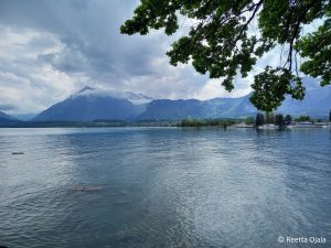
The location by the Lake Thun was beautiful.
We arrived in Thun a day before the scientific program started, so we had some time to adjust and to see the surroundings a bit. This turned out to be a good decision, as the conference days were long and tiring, because there were so many interesting topics; transformation of sensory evidence into action, what role do emotions have between sensation and action, large-scale neuronal computations and much more.
At first, I felt overwhelmed in the middle of all the socializing action. Luckily, I was not there alone, and my colleague dragged me to meet other people. We are both starting our PhD projects in the same lab soon and we got to know other PhD students that are a little further in their studies. I ended up having very vivid conversation with one young woman about animal research ethics, which is very close to my heart. In the end I exchanged contact information with quite a few other students too.
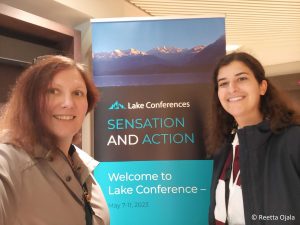
Me and my colleague Joana Doutel Figueira
In the last evening, it was time to face my biggest fear, the poster presentation. It started in the worst possible way. A big name in the field came to hear me. I felt my cheeks turn red and I started to stammer. I took a deep breath, gathered myself and started talking more slowly. A little voice in the back of my brain was shouting that this person knows so much more than me; how should I present all this? But I remembered my PI’s words. This is my work, and I am the expert on it. So, I just explained everything, some parts of the analysis more briefly. I think the professor saw my struggle. He was very friendly and made some very good questions and gave constructive feedback. We talked for a while and after he left, I wrote down the comments to go through them later. After that it was much easier to present to the other students.
I am very happy that I had this opportunity. And I am proud of myself for overcoming my fears and talking to people. I got new ideas for my project and new perspectives on this area of research. The scientific talks and the conversations with others made me feel that I am on the right path in my life. I want to encourage all the students to apply to conferences! The information load will be heavy, but the point is not to take every talk as a lecture. It is more about getting inspiration and making connections, meeting other people who are interested in the same things that you are. I surprised myself positively, but there is still much to learn. Maybe next time I dare to go to talk to the PI whose talk I found totally fascinating.
Have you ever stared closely at fish in an aquarium? Isn’t it just amazing to look at the plethora of shapes, colors, and different behaviors that the fish, living on the other side of the glass, display? My younger self really enjoyed the wonders of the aquarium hobby – I believe the underwater world bears some of the world’s most amazing representations of life, and those tanks could, in some way (ethical issues aside), depict a little part of these marvels. But, how do fish see each other? Are they attracted to the colors other fish display? If so, what happens then, in the dark?
I am Adrián Colino Barea, a Spanish wildlife biologist, and a current first year student of the Master’s Programme in Ecology and Evolutionary Biology at the University of Helsinki. My main motivation to join this programme here was to dig deeper in applied ecology in tropical regions, as the University of Helsinki has several research lines in the tropics. I am aiming to focus on biodiversity conservation: I want to contribute and make a difference to revert the trends that current human-driven impacts have on nature globally.
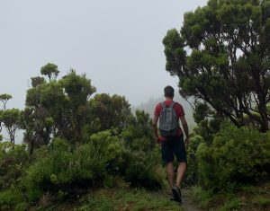
Not surprisingly, me in the outdoors, hiking, looking for some interesting endemics in Azores. Greatly attracted by field ecology, I would not have pictured myself doing this exciting lab-based internship back when this photo was taken!
While my passion has always been working on the field, touring the fish room of the Integrative Evolutionary Biology (IntEvoBio) lab during an introductory course of the master’s last year awakened my younger self thrill. Racks with tens of tanks full of cichlid fish from the African Great Lakes fill the room, all around us, while some of the IntEvoBio lab members, led by Prof. Claudius Kratochwil, guided us through the nuts and bolts of their interesting job with all these fish, and why they are so particularly wonderful.
African cichlids: the book example of speciation
The African Great Lakes are three massive freshwater bodies in tropical West Africa, originated by ancient tectonic shifts. However, these lakes have borne livable conditions for a relatively short time. A certain group of cichlid fish got to these waters and started evolving at an unprecedented rate, fitting every possible niche, and generating entire, complex ecosystems, all based in different cichlids. What is more, they display very different color patterns of colors, bands, and spots, and perform elaborated mating rituals. Their unusually fast evolutionary process is based in ‘sexual selection’, meaning that individuals choose others as partners based on certain traits of their preference, despite the fact that these traits could make them, for instance, more showy – and therefore easier to spot by predators, hence against ‘natural selection’ as described by Charles Darwin.
To put it in perspective, for any evolutionary biologist, the cichlids of the African Great Lakes are old acquaintances, and for some they are the entire reason for their research. For many biologists, they are just inexplicably special. Some even say that the basis of evolution today would be radically different, had Darwin visited these lakes instead of the Galápagos Islands almost 200 years ago, and looked to these fish.
While in the lab, I enjoyed seeing all of the complexity that I have read about, right before my eyes. The researchers explained to us how these fish (and their evolutionary process) strongly rely on color communication. They have developed color signaling to inform about their sexual status, the hierarchy within a group, and even some species can change their colors rapidly, to give fast information!
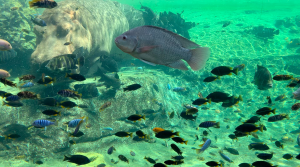
Cichlids have evolved into a big variety of colors, shapes, and behaviors among the waters of the three African Great Lakes. Just to clarify, the big guy on top left is not a cichlid 😉
However, one of the drawbacks to this very fast evolutionary process is that it is also very fragile, as species may seem radically different in colors and shapes but have very similar DNA, because of how recently they have evolved. In addition to this potentially fragile evolution, these lakes are particularly vulnerable to the impacts of human development. Lake Victoria is the second-largest lake on the planet by area and a huge population increase, has led to 40 million people now inhabiting the coastal region This human activity has resulted in major issues related to biodiversity, mostly through pollution, overfishing, and introduction of invasive predator fish and floating plants.
How does sexual selection work under deterred underwater light conditions?
In particular, an invasive floating plant, the water hyacinth (Pontederia crassipes), is causing terrible damage to all ecosystems in Lake Victoria, and migh spread to other lakes. These plants prevent light from penetrating, hindering the oxygen production in the water and making the ecosystems within it collapse. Moreover, the human communities around the lake cannot physically access their resources because of how dense and inexpugnable the floating mats are.
But, coming back to my initial concern, floating plants – as well as increases in turbidity – make the underwater dark. Is there anything happening directly to the cichlids? If they rely so strongly on visual communication for their daily lives and long-term evolutionary process, how do they cope under a scenario in which their vision – light supply – is hindered? How can they find and recognize each other effectively and ‘sexually’ select their preferred partners according to visual signals (colors)?
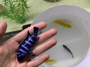
During my internship, beyond my research project, I am learning the basics of tank care and manipulating fish. I certainly work with beautiful animals, such as this Pseudotropheus (Chindongo) demasoni, and the yellow females of Ps. (Ch.) saulosi in the bucket.
Back during my lab visit, I asked some researchers and they didn’t have an answer. This visit was inspiring enough to raise meaningful biological questions, with profound biodiversity conservation meanings. I am very pleased that HiLIFE gave me the chance to shed light on this topic, by granting me a Research Trainee Scholarship. Since March, I can call Prof. Claudius Kratochwil my supervisor, and some of the PhD students that once showed me through their research, my helpful mentors.
From March to today: the beginning of my experimental phase
During these months, I have been setting up my experimental arena, and dedicatedly come up with a protocol to follow. I am working on the mate choice of cichlid males over females under different light conditions. Only last week I started the experimental phase, and I am excited to know what will happen with the results that I am obtaining – I will keep you posted on my progress. If this experiment arise meaningful outcomes, these invasive floating plants would potentially be given a new threat to nature, and hopefully a new reason to focus efforts and find an effective solution in the field, for the cichlids, for the people in the region, and for the entire ecosystems.
Hi fellow students! My name is Celia Gómez Sánchez, and I am a second-year master’s student in Genetics and Molecular Biosciences. In October 2021 I received the opportunity to attend the American Society of Human Genetics (ASHG) 2021 annual meeting. The conference was held in the US with a hybrid model, so attendees could watch the talks either on-site or online. This enabled people from all over the world to participate ¬–including me. Because there were multiple events happening simultaneously, during the week when the ASHG21 meeting happened I only attended the talks I was most interested in; nevertheless, the conferences were recorded, so they were later accessible to everybody like me that could not watch them live. This was a great feature since I was very keen on most of the topics, and wanted to make the most of this opportunity 😀
As a master’s student, I didn’t present any type of research, I only listened to the conferences. However, this was enough to learn a lot of about various topics, some of which were closely related to my master’s thesis that I’m currently working in. I was especially interested in genetics and neurosciences, and the talks concerning neurodevelopmental diseases were definitely my favorites. We learnt about the use of both cells and mice as models for the different disorders, and how they had explored the genome to find disease-causing variants. For example, one of the groups were able to identify autosomal recessive variants in genes previously unlinked to autism spectrum disorder, resulting in 31% rate of patient diagnosis. Furthermore, the talks explained the use of genomic techniques that I had recently studied (such as single-cell ATAC-seq or single-cell transcriptomics), which provided me with a great opportunity to understand better the application of these techniques with real examples and the results they provide with.
Moreover, some of the meetings specifically focused on craniofacial development, my area of research at the moment. I am studying the cranial neural crest (that gives rise to the craniofacial region), and certain conditions related to defects arising from it, such as pituitary hormone deficiency and maternally inherited gingival fibromatosis. These conditions result in delayed growth and puberty and craniofacial malformations. What is very interesting about the neural crest is that 30% of all congenital malformations in humans are derived from abnormalities in it, since the neural crest precedes the formation of multiple tissues in the embryo. Therefore, its study is key to preventing and treating multiple birth defects, and so I was very excited to learn about state-of-the-art research involving this embryonic structure. As a matter of example, I was glad to hear about TFAP2A, a cranial neural crest marker on which I have been focusing my experiments. I learnt that enhancer mutations that dysregulate this gene were found to cause branchio-oculo-facial syndrome (BOFS), a condition that results in eyes and ears malformations together with characteristic facial features.
In conclusion, being able to attend the ASHG21 meeting was a very powerful experience, that allowed me to learn about multiple topics of interest and get in touch with current methods to research human diseases. I am very happy to have been able to attend these conferences and I really thank HiLIFE for this opportunity, that I wouldn’t have been able to get on my own.
Hi, and welcome to the HiLIFE-trainee blog! My name is Akseli Bonsdorff and I am a fourth-year medical student at the University of Helsinki and also working on my PhD-thesis in pancreas transplantation and general pancreatic surgery. My first scientific article on the role of early plasma amylase levels in predicting pancreas graft-related complications after pancreas transplantation was published in early 2021 and I received an opportunity to present the key findings of the study at the European Society of Transplantation (ESOT) 2021 congress. ESOT is a biannual congress that brings together transplantation surgeons, nephrologists, hepatologists, pathologists, nurses, and students – such as me – and acts as a platform for discussing novel topics and state-of-the-art findings in the field of transplantation.
This year, ESOT was held as a hybrid conference, and attendees had the chance to choose between on-site or online attendance. For approximately ten to twelve hours a day, I sat and listened through the many inspirational talks, keynotes, presentations, and discussions on topics ranging from the definition of brain death to the implementation of artificial intelligence applications in transplant organ allocation systems, and from the effect of Covid19 on the transplant communities to why living donor liver transplantation is not performed extensively in Western countries but cover the majority of liver transplantations in Asian countries. I am still dumbfounded by the exhaustive coverage of different topics, and probably still- after two weeks from the congress – processing all the new knowledge I gained.
On the third day of the congress came the moment of my presentation. Due to the hybrid model, I had to prerecord the bulk of my presentation and my role during the session was to answer questions arising from the audience on-site and online. The two dutch professors chairing my session were not too harsh on me, and one of them even thanked me for an interesting presentation (which he did for every presenter, but needless to say, it felt nice at the moment). Hybrid or not, my debut was something I will remember for a long time.
Without a doubt, ESOT2021 had provided me everything I expected and probably even more. I had the chance to delve deeper into my own field of research (that being pancreas transplantation), but also received tasters into the other subspecialties in transplantation. I am extremely grateful for the financial and scholarly help from my great mentors Adj. Prof. Ville Sallinen and Adj. Prof. Ilkka Helanterä. I also thank HiLIFE for the support received. Making attending an event like this possible for students and aspiring researchers is extremely appreciated.
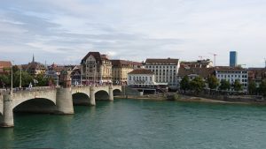
My name is Teemu Kuosmanen and I am here in Basel for my master’s thesis research which focuses on the exciting and relatively novel field of mathematical oncology.
Cancer is conventionally seen as a genetic disease and characterized by the accumulation of genetic and epigenetic alterations. While this is of course per se true, such definition naturally implies that the focus of cancer research should be in the systematic study of mutations and genes. Indeed, this has and continues to be the central dogma and interest of mainstream cancer research. Continue reading “Greetings from Basel, Switzerland”