Plants sprouting in my garden as well as in my computer – Experiences during my HiLIFE Traineeship


My four-month HiLIFE traineeship spanning from furious snowstorms followed by a tender spring awakening to 19 hours of sunshine a day has now come to an end. Not only Helsinki’s residents had to endure extreme temperatures, the plants in my models were also challenged by various climates. In short, I modeled the climate induced species range shift of the flowering plant St. John’s wort (Hypericum perforatum) using a dynamic eco-evolutionary modeling approach. In long, check out my previous Blog Post “Gathering the puzzle pieces for my simulation – The beginning of my HiLIFE traineeship (10th April 2024)”.

And what a journey it has been! I remember precisely how vague the picture of my modeling project seemed during the start of my internship. I crawled through a virtual pile of papers, data, models and ideas. I was wondering how all of that fits together. But every day the picture became more and more clear. And indeed, by the end of my internship I managed to combine all the puzzle pieces and build a shiny simulation. Hurray! At this point: Many thanks to my supervisor Dr Frédéric Guillaume (University of Helsinki) for the support!

My plants growing happily in my laptop… or more precisely on the supercomputer Puhti. The picture is AI generated with Canvas Magic Media.

But how did I manage to get there?

I’m not gonna lie: It was a roller-coaster ride! Shortly after the start I found myself rolling slowly but surely towards the first steep slope: species distribution modeling. Suddenly, scary terms like Random Forest, True Skill Statistic, Pseudo-Absence, and Ensemble Forecast became part of my vocabulary. But it paid off: Fitting a super fancy species distribution model was a true downhill ride (including loopings). Fun! From this distribution model I could derive the occurrence probability of my plants based on the temperature in each location in Finland. Hm, but climate is not the only factor determining species distribution. Land cover also plays a role, or have you ever seen a flower growing on a lake? Me neither. So, I filtered out all the unsuitable locations in Finland.

Maybe you remember this quirky looking map from my first blog post showing the average temperature in January and the occurrence of my plants in Finland… Well, luckily, I managed to use these data to fit a proper species distribution model. Tadaa! Doesn’t the new map look fancy? The plots were generated in R Studio with the packages terra and biomod2.

What’s next? The roller-coaster was already taking off for the next hill to climb: statistically analysing the effect of temperature on characteristics like number of flowers and seed weight. Or in other words, clearing my way through the jungle of Reciprocal Transplant, Resurrection and Greenhouse experiment data (kindly provided by Dr Marko Hyvärinen and Dr Maria Hällfors from LUOMUS, UH, and SYKE). My best weapons were quadratic log gaussian model and negative binomial hurdle model. With those I was able to understand how sensitive my plants are towards temperature. Another puzzle piece!

Finally, I was approaching the last bit of this wild roller-coaster ride. Essentially, I was spiraling down towards plugging everything into nemo-age, the simulation modeling tool developed by Dr Guillaume and colleagues. One key aspect here was to include the genetic basics of evolution to allow my plants to adapt to changing temperatures – the heart of a dynamic eco-evolutionary modeling approach. Piecing the puzzle together included a lot of data formatting and tweaking around but I’ll spare you the details. I’d rather join you on a shortcut towards the results of my simulation.

How are the plants doing?

According to my simulations they were doing good and would do even better in the future. That means, the distribution range was predicted to expand north with increasing temperatures. That is quite logical as my data analyses revealed that the St. John’s wort performs better in warmer climates. Additionally, the plants were adapting to the warming climate. This fits well into the general picture that warm adapted plants are predicted to expand northwards (in the northern hemisphere), while cold adapted plants slowly lose their habitat if they are not able to adapt.

A piece of Finland, longitude-wise spanning the whole country and latitude-wise spanning from Lahti to Tampere, approx. The coloured background represents the average temperature of the warmest quarter, and the black dots represent the simulated St. John’s wort distribution. Time is increasing from the top-left to the bottom-right (2001-2090). The plots were generated in R Studio with the base package.

So, the simulations confirmed the often-predicted range expansion of warm adapted plants towards the north. The role of adaptive evolution could further be investigated by modifying the evolutionary parameters in the model. But the story of my HiLIFE traineeship ends here. As one might say, all’s well that ends well. The roller-coaster ride as well as the simulations ran through successfully. The flowers in my simulations and in my garden have sprouted. This ride has been a real pleasure and I hope you enjoyed it as much as I did.

– Theresa Koller

The plants seem to do great and enjoy temperatures that are a bit warmer than the historical cool Finnish climate. Credits for the idea to use this meme: Nicholas Armeni. Source: pinterest

Discovering the potential of next-generation probiotics (NGPs) as a HiLIFE trainee


I’m Pinja Perkkiö, an aspiring microbiologist and a HiLIFE scholarship trainee in Reetta Satokari’s lab (University of Helsinki, Faculty of Medicine)! I’ve just finished my bachelor’s degree and I will start my studies in the master’s programme of Microbiology and Microbial biotechnology next autumn. This summer, I will be diving deep into the world of gut microbiota as I research the microbiota restoring potential of NGPs in intestinal inflammation and dysbiosis. 


How antibiotics disturb our gut microbiota 

The gut microbiota is defined as a community of microbes, e.g. bacteria, fungi, viruses and archaea residing within the gut. The microbiota is responsible for many important functions, such as breaking down complex carbohydrates and protecting the gut from infections. When a person takes antibiotics, it not only kills the bacteria causing the disease, but also the beneficial ones as antibiotics aren’t strain-specific. This allows potentially pathogenic bacteria to grow in larger numbers – the situation is called dysbiosis, meaning that the microbial community composition has been altered in a harmful way. 

Luckily, we have a tight layer of cells – the epithelium – that lines the walls of the gut and is covered by two layers of mucus. The mucus combined with the epithelium acts as a barrier between our intestines and the rest of our body, preventing intestinal pathogens from causing too much harm. Normally, it is very difficult for microbes to cross the mucus layer as it traps microbes and is loaded with antibacterial substances. In addition, the cells of the epithelium are tightly attached to each other, preventing any further movement of microbes. There are also lots of immune cells embedded in gut tissues that are ready for action, should a pathogen try to cause an infection. 

However, as dysbiosis leads to the growth of pathogenic bacteria, our immune cells (neutrophils, NK cells and macrophages, for example) activate inflammatory reactions. While these reactions are necessary for the elimination of pathogens, they also damage gut tissue. Pathogenic bacteria also activate the secretion of inflammatory molecules, cytokines, in a type of gut epithelial cell called enterocyte, which leads to the epithelium getting “leaky” – this means that microbes can cross the epithelium more easily and cause further inflammation in gut tissue. These bacteria can not only cause nasty infections, but they may also travel deeper in gut tissue or even to other sites in the body, increasing the risk of certain cancers (Bastos et al., 2023; Kouzu et al., 2022)! 


Next-generation probiotics against dysbiosis 

To fight antibiotic-induced dysbiosis and reduce the inflammation and leaky gut syndrome caused by it, researchers have turned their attention to potential new bacterial strains that could function as next-generation probiotics. They are defined as “live microorganisms identified on the basis of comparative microbiota analyses that, when administered in adequate amounts, confer a health benefit on the host” (Martín R, Langella P., 2019) – in simpler terms, microbes that can potentially help boost our health. Traditional probiotics, such as Bifidobacteria and Lactobacilli, have shown great potential in restoring a healthy microbial community in the gut after taking antibiotics, thus preventing dysbiosis and infections. They have also been shown to inhibit the production of inflammatory molecules in both immune cells and epithelium and boost the production of anti-inflammatory molecules.  


Probiotics exert anti-inflammatory effects on several cell types. Created with Biorender.com. 


There’s still lots of research to be done to bring next-generation probiotics into wide use. More information is needed on the different strains of potential probiotic bacteria and their specific effects on both the immune system and the gut epithelium. This is where my research project comes in! 

The project I’m collaborating on focuses on a mix of 14 different anaerobic bacterial strains that have the potential to restore the gut microbiota after antibiotic use and prevent or reduce inflammation. The next-generation probiotic mix was originally tested in an in vitro mucosal-simulator of the human gastrointestinal tract (M-SHIME(R)) model. A natural gut microbiota was obtained from the stool sample of a healthy human donor, after which it could grow. Then, the “gut” was exposed to antibiotics to induce dysbiosis. After the antibiotic treatment, one system was treated with the probiotic mixture and the other one with a negative control. Finally, samples from probiotic- and control-treated microbiotas were taken. Bacteria present on these will be studied to check the impact of the antibiotic intake in the microbiota and compare its possible recovery after the probiotic treatment. The production of short-chained and branched fatty acids, lactate and ammonia at different time points will also be measured since their levels indicate how much beneficial bacteria there are compared to harmful ones. 

To study how the anaerobic isolates might affect intestinal inflammation, I will culture two different intestinal epithelial cell lines: Caco-2 and HT-29. These cell lines were chosen because they are well-characterized and resemble enterocytes on the gut epithelium. Then, I will expose the HT-29 cell line to either the anaerobic bacterial blend or a placebo. After incubation, pro-inflammatory IL-8 production in the HT-29 cell line will be measured using ELISA assay. Later, the capability of a single freeze-dried strain in attenuating the IL-8 levels produced by HT-29 cell line will be assessed.  

Caco-2 cell line will be used to measure the effect of the microbiota samples collected from M-SHIME(R) on the barrier integrity of the Caco-2 (enterocyte) monolayer. The integrity will be assessed by measuring the TEER (transepithelial/transendothelial electrical resistance) of the cell monolayer, and the results will tell us if our strains can alleviate leakiness in the epithelial cell layer caused by dysbiosis. 


HT-29 cells under a microscope. 


Starting my research 

So far, I’ve read lots of literature related to the gut microbiota and treating inflammation with probiotics and next-generation probiotics. This has given me a good foundation on which to build my knowledge. The steps using M-SHIME(R) human gut simulator have been completed previously, so I’ve mainly been growing the intestinal cell lines required for our experiments. Luckily, I already have some experience with cell culture, so the start hasn’t been too difficult. My group has also been supportive and helpful, so I’m optimistic about the upcoming months! 


Seeding HT-29 cells for our experiments. 


The next step will be starting the IL-8 induction experiments, and once the initial results are ready, we can proceed to plan further. If you’re interested in our results, stay tuned for my next blog post! 


Sources: 

Bastos AR, Pereira-Marques J, Ferreira RM, Figueiredo C. Harnessing the Microbiome to Reduce Pancreatic Cancer Burden. Cancers (Basel). 2023 May 5;15(9):2629. doi: 10.3390/cancers15092629. PMID: 37174095; PMCID: PMC10177253. 

Kouzu K, Tsujimoto H, Kishi Y, Ueno H, Shinomiya N. Bacterial Translocation in Gastrointestinal Cancers and Cancer Treatment. Biomedicines. 2022 Feb 4;10(2):380. doi: 10.3390/biomedicines10020380. PMID: 35203589; PMCID: PMC8962358. 

Martín R, Langella P. Emerging Health Concepts in the Probiotics Field: Streamlining the Definitions. Front Microbiol. 2019 May 21;10:1047. doi: 10.3389/fmicb.2019.01047. PMID: 31164874; PMCID: PMC6536656. 


Some literature: 

Berta Bosch, Saliha Moutaharrik, Andrea Gazzaniga, Kaisa Hiippala, Hélder A. Santos, Alessandra Maroni, Reetta Satokari. Development of a time-dependent oral colon delivery system of anaerobic Odoribacter splanchnicus for bacteriotherapy. European Journal of Pharmaceutics and Biopharmaceutics. 2023; 190. 73-80. https://doi.org/10.1016/j.ejpb.2023.07.010. 

Cristofori F, Dargenio VN, Dargenio C, Miniello VL, Barone M, Francavilla R. Anti-Inflammatory and Immunomodulatory Effects of Probiotics in Gut Inflammation: A Door to the Body. Front Immunol. 2021; 26(12):578386. doi: 10.3389/fimmu.2021.578386.  

Marzorati M, Van den Abbeele P, Bubeck SS, Bayne T, Krishnan K, Young A, Mehta D, DeSouza A. Bacillus subtilis HU58 and Bacillus coagulans SC208 Probiotics Reduced the Effects of Antibiotic-Induced Gut Microbiome Dysbiosis in an M-SHIME® Model. Microorganisms. 2020; 8(7):1028. https://doi.org/10.3390/microorganisms8071028 

How my experience as a HiLIFE trainee brought out the bookworm inside me

My HiLIFE traineeship has come to an end, and although it is sad to close a chapter, I couldn’t be more excited to share how my experience went.

I am Mireia Pagès Guitart, I just graduated with an MSc in Pharmaceutical Research, Development, and Safety at the University of Helsinki. In September 2023, I started an incredible project at Michael Jeltsch’s lab to tackle a huge sustainability challenge with one of the best tools at our disposal: science. You can learn more about it by reading my previous post, by clicking here. In January 2024, HiLIFE supported my investigation by awarding me the HiLIFE research traineeship. Do you wonder how it ended up? Keep reading!

After months of reading, testing, failing, achieving and, most importantly, having fun, I am happy to share that I succeeded in expressing GFP in vitro. Unexpectedly, it turned out that earthworms have inherent green fluorescence. Therefore we could not be sure whether the green color after in vivo imaging was due to our protein or due to the animal. Even so, my project was a big step forward to achieve our goal: developing a sustainable animal model for protein production and microplastic degradation. You might wonder: what now? I will continue my journey in Catalonia (for now) and my lab team will continue this amazing project… so be aware of a promising future!

Worm Imaging. How cool is it?

The HiLIFE traineeship made me realize I enjoy research more than I thought! Doing extra hours, having to check up on the cells during weekends, and messing up experiments doesn’t matter if you have science at your heart. During my experience, I thought about the thin line between a job and a hobby, and how my project became my pastime. Feeling this way towards research couldn’t have been possible without being surrounded by a comforting team. I am aware of how lucky I was to find a lab that matched my energy, sense of humor and enthusiasm for research.

Persian dinner by my colleague Honey, with my lab group. So delicious…

Joining a research group goes beyond the lab work. I was proud to join workshops, competitions, conferences and talks that were as important to grow as a scientist. The value of grasping every opportunity during my stay brought out the bookworm inside me. Nothing but good outcomes in my research and personal progress came afterwards. I encourage future HiLIFE trainees to make the most out of their traineeship.

Our lab team as “The Bunsen Burners” for the Research Program Unit competition 🙂

Over my master’s at the University of Helsinki and my stay at the lab, I have internalized many learning lessons from which I want to share three:

  1. Whatever you do, stay curious and skeptical, and, above all, enjoy the journey because there is no other destination (by my PI Michael).
  2. Sometimes you think you have been buried, but actually, you have been planted (by the activist Christine Caine)
  3. Surround yourself with people who are different from you: open your eyes and envision your view of this small world with big souls (by my friend Achmet).
Spring in Helsinki looks nice!

A chapter closes and another begins. Farewells are sad, but they remind you of the people that you have met along the way, and that you can proudly call them friends. They remind you of your social impact on new environments, and how people learn from you as much as you gain from others. Farewells are not a goodbye, but a “see you later”!

In catalan I would say: aquesta experiència no ha sigut bufar i fer ampolles, però res extraordinari ho és! – this experience hasn’t been easy, but nothing extraordinary is. 

On dinoflagellates, methane and ice floes: the beginning of my internship

What can you do when you are supposed to go to sea to sample plankton and you notice in the morning that a great amount of ice has floated in to block the harbour? Well, go slowly and carefully and prepare for a workout and some cold fingers. And how do the changes in phytoplankton communities affect the global methane budget? I guess that’s what we want to research.  

Hi, I am Evert Odé, a second-year Bachelor’s student in Environmental Sciences at the University of Helsinki and one of the recipients of this year’s HiLIFE Research Trainee Scholarships. I am especially interested in marine ecosystems and their interactions with the Earth and climate systems in these agonising times of great human-induced changes. Phytoplankton, those tiny organisms at the base of most marine food webs, are at the centre of these interactions: they affect the climate system by sequestering carbon dioxide and influencing the formation of clouds but are also affected by climate change.  

It is well known that phytoplankton constitute an important carbon sink, and people have even proposed that the oceans should be fertilised with iron to fight climate change. However, phytoplankton can also emit greenhouse gases. This all makes the interaction between phytoplankton and climate change a very fascinating subject indeed, and I am glad that I have the opportunity to conduct my four-month internship in the Plankton Biodiversity Research group at Tvärminne Zoological Station and participate in their research on this matter.  

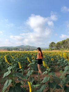
Me and a nice view of the archipelago off Tvärminne in early March

One topic we are especially interested in studying is methane production by different kinds of phytoplankton. For a long time, it has been a paradigm that biotic methane production only happens in environments where oxygen is not present1, such as sediments and bogs rich in decaying organic matter, and the digestive systems of ruminant animals. Moreover, only certain types of Archaea, those intriguing members of the third domain of life, have been considered capable of methane production2: the methanogens, which inhabit these anoxic environments. However, there have been signs that this standpoint probably doesn’t explain all methane production that is being detected. Especially the phenomenon called the “marine methane paradox” has been challenging to explain from this point of view: methane concentrations in oceanic surface waters are in supersaturation relative to the atmosphere (that is, they contain more methane than they would if they would be in balance with the atmospheric concentrations), even though they are rich in oxygen and far away from the potentially anoxic sediments1.

In recent decades, the paradigm of methanogens as the only methane producers has begun to crumble, as an accumulating number of studies have revealed that many organisms in both Eukaryote and Bacteria domains do produce methane, even though the exact pathways are not always clear. For us, the most interesting aspect of this new research is that a number of phytoplankton species have been demonstrated to produce methane, and even in association with photosynthesis, which produces oxygen! Since oxygen (a very oxidising substance, as could be concluded from the name) could be considered an enemy of methane, a reduced hydrocarbon substance that has been thought to form only in anoxic conditions, this is quite a counterintuitive finding. It also illustrates that one should be cautious with paradigms, especially in the field of microbiology.  

The most important aspect of all this might be how these new findings affect the global methane budget. Even though the methane production by phytoplankton and other organisms is a natural phenomenon that has been going on for millions of years, it can significantly affect the calculations of global emissions and sinks of methane3. This could, in turn, affect our thoughts concerning significance and quantification of other, more human-influenced sources of this potent greenhouse gas. In addition, phytoplankton-derived methane emissions can also change notably4 due to the great anthropogenic changes of our time.  

The phytoplankton community consists of many groups of organisms with very distinct evolutionary backgrounds and widely differing traits. Cyanobacteria, those ancient blue-greenish organisms that have the honour to be the evolutionary innovators of oxygenic photosynthesis, have been shown to produce methane2. Their summer blooms have become a common phenomenon in the eutrophicated and warming Baltic Sea, as well as in many lakes (a well-known nuisance to many enjoyers of aquatic environments), and in our project, we want to further investigate the climatic effects of their increasing dominance. Their competitors among the summer phytoplankton include the cryptophytes which we are also interested in researching. 

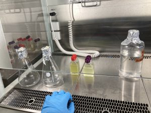
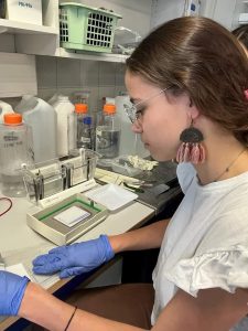
Starting to inoculate some algae for our experiments (in this case Synechococcus sp., very small cyanobacteria)

Probably an even more interesting group of phytoplankton from our point of view are the dinoflagellates. They are indeed strange creatures: many of them photosynthesize with chloroplasts acquired through secondary, tertiary and even quaternary endosymbiosis (which means that their ancestors have engulfed an alga, whose ancestors have engulfed an alga, whose ancestors have engulfed an alga, whose ancestors have engulfed a cyanobacterium… crazy, isn’t it?), but they can also eat other organisms. And move around the sea by whirling their flagella! 

Methane production by dinoflagellates has not been researched much, even though they are increasing in abundance in many areas, including the Baltic Sea. Their increase here during the phytoplankton spring bloom has come at the expense of diatoms. Diatoms are another, still more common and diverse group of phytoplankton. They do not eat anybody or move around much, but they are quite efficient in photosynthesis and nutrient uptake, and their surface is made of glass (or very nearly so). In the upcoming experiment this spring, we will investigate the effects of the dinoflagellates, diatoms, cyanobacteria and cryptophytes on the sequestration and release of methane and carbon dioxide.  

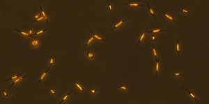
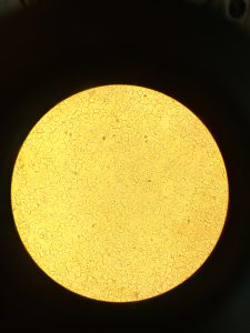
Cells of Phaeodactylum tricornutum, a diatom species that we are using in our experiment, with two different morphological types visible in the picture

Well, what about the struggle against the ice floes? In addition to laboratory experiments, our project also includes field monitoring and measurements. The ice situation at the beginning of March was difficult due to the fast ice surrounding the harbour. When the fast ice finally broke, we decided to sample on next Thursday morning. On Wednesday, I watched from the window of my microscopying room as the countless ice floes floated slowly from the sea to the harbour driven by some change of wind. It didn’t seem very promising for our field work. 

The next morning, the drift ice was still there, blocking the harbour, even though the waters further out were quite ice-free. We still obtained a permit to try; but very slowly… So, we boarded Crangon, an 8,2–metre–long research vessel, and started a long and arduous journey through the drift ice. Research technician Kurt Spencer drove the boat, while postdoc researcher Per Hedberg kept the ice floes away from the propeller with a boathook and I tried to push ice aside from our route at the front of the boat. It took an hour for us to reach the open water not so far away, but we were very satisfied when finally getting to take our samples. This was done quite quickly and then… all the way back again. Hopefully this would not take much longer, for then we would miss lunch.  

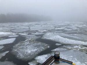
The drift ice that we had to struggle through (the weather was foggy, which added nicely to the ambience of the scene).

So, we started pushing ice diligently once again. I was unsatisfied with my telescopic boathook pole, which clearly was not designed to be used for this purpose and didn’t stay in its length when pushing the ice floes away. When we started returning to the harbour through drift ice, I saw a long piece of driftwood, probably a fugitive lath from some faraway building site. I picked it up from the icy sea with the boathook, and it turned out to be a useful tool for ice manipulation. Sometimes, driving through a combination of many large ice floes was quite a puzzle, but in the end, we managed to make it back by lunch. Even though my fingers were quite cold from sampling and handling the wet piece of wood, I thought it was a fun way of doing science.  


Some literature:

1Bižić, M., Grossart, H.-P. and Ionescu, D. (2020) Methane Paradox, In eLS, John Wiley & Sons, Ltd (Ed.). https://doi.o rg/10.1002/9780470015902.a0028892. 

2Bižić, M. et al. (2020) Aquatic and terrestrial cyanobacteria produce methane. Science advances. [Online] 6 (3), eaax5343–eaax5343. 

3Günthel, M. et al. (2019) Contribution of oxic methane production to surface methane emission in lakes and its global importance. Nature communications. [Online] 10 (1), 5497–10. 

4Klintzsch, T. et al. (2020) Effects of Temperature and Light on Methane Production of Widespread Marine Phytoplankton. Journal of geophysical research. Biogeosciences. [Online] 125 (9). 


Gathering the puzzle pieces for my simulation – The beginning of my HiLIFE traineeship

Hi! I’m Theresa, a HiLIFE Trainee 2024, and I’m now in my fifth week of my traineeship in the Eco-Evolutionary Dynamics Lab by Frédéric Guillaume (UH, OEB), in collaboration with Anniina Mattila from LUOMUS. The goal of my project is to predict the species’ range shift of the St. John’s wort (Hypericum perforatum) across Finland following climate change. That means I want to know in which places the St. John’s wort will be able to persist, in which places it will go locally extinct and which places it will newly colonize. As the temperatures in Finland are going to increase, I expect the St. John’s wort populations to move north since the climate is cooler in nothern latitudes. However, it is not that easy because species’ responses to climate change depend on many different factors. And that is exactly why I will use computer simulations!

The beautiful St. John’s wort (Hypericum perforatum). Picture is taken from https://gobotany.nativeplanttrust.org/.

But how did I end up choosing this project?

I’m a first year Master’s Student in Ecology and Evolutionary Biology at the University of Helsinki. I’m originally from Munich, Germany, and I did my Bachelor’s in Molecular Ecosystem Sciences at the University of Göttingen, Germany. During that I discovered ecological modeling and loved it because I enjoy programming and describing nature with math. For my B.Sc. thesis I modeled bird diversity in an agricultural system. I also started to be very fascinated about evolution and genetics, with which I came in touch during my Bachelor’s but never had the opportunity to work with it. Until now! Early in my Master’s at the University of Helsinki I found the Eco-Evolutionary Dynamics Group, which combines modeling with the genetic mechanisms behind evolution. It’s a match!

How will I be able to predict the future?

I must admit, that’s a very bold statement and of course I’m not a clairvoyant. But putting some assumptions and limitations aside, such simulations are indeed very powerful in forecasting the future, or at least make really good guesses. So how will I make really good guesses? The answer is: A dynamic eco-evolutionary modeling approach, that is based on individual-based genetically and spatially explicit simulations, which in turn are calibrated with empirical data. But what on earth does that even mean?

A dynamic eco-evolutionary modeling approach allows a more integral way of describing species’ responses to a changing environment than most other modeling approaches. The magic behind it is that it includes 1. the ability of a population to colonize new areas with more favorable climatic conditions and 2. its capacity to genetically adapt to new conditions.

This approach will be realized by running individual-based genetically and spatially explicit simulations using Nemo, a tool developed by the Guillame Lab. Nemo simulates each plant individually including its genetic architecture, life cycle and location. And that gives the possibility to include evolutionary as well as ecological key processes such as mutation, genetic drift, selection, and dispersal.

These simulations take in many different parameters and this is where the empirical data comes into the equation. As I want my simulation to represent reality as accurately as possible, I will extract the needed parameters from real-world data, such as climate data, species distribution data, genetic data and thermal performance data. Then, I will plug in those parameters into the simulation together with IPCC climate change scenarios and observe what happens. Exciting!

Here you can see a map of Finland, together with the monthly average temperature (1970-2000) in January across Finland (color coded) and the occurrences of St. John’s wort (black dots). This is just a preliminary visualization of some parts of the data I will use. Climate Data was taken from WorldClim, Species Occurrence Data was taken from GBIF, the plot was made in R, with the terra package.

Where am I at?

I spend the last few weeks geting a grasp of what I need for my simulation, how I will get it and how everything is connected. Now as I have a plan, the first thing to do is relating current climate data to the current species distribution. That will help me getting an idea which climatic conditions are suitable for the St. John’s wort. And that in turn is one of the very important parameters needed in my simulation. In the upcoming months I will analyze many more data sets until I can feed the simulation with real-world paramters and finally answer the question: How will the finnish St. John’s wort populations do in the future?

Earthworms as the ultimate strategy to face microplastic pollution

Have you ever seen an earthworm make magic? These little creatures can, for instance, eat leaves and make them disappear! Earthworms are one of the few animals capable of decomposing organic material. So, even though they seem insignificant living things, it is hard to imagine a world without them. But, what if I told you earthworms could also make microplastics in soil vanish? What if they represent the ultimate strategy to face microplastic pollution?

I am Mireia Pagès Guitart, a Catalan biomedical scientist, a second-year student of the Master’s Programme in Pharmaceutical Research, Development and Safety at the University of Helsinki, and a new HiLIFE trainee! With the support of the HiLIFE Research Trainee Scholarship, my ambition was to engage in applying sustainability to science, a much-needed climate action.

Me working in the lab, performing a Western Blot. If I had known what my future research would be like back then, I don’t know if I would have believed it!

Even though my passion has always relied on combining both topics (sustainable science), this dream didn’t come true until I came upon Prof. Michael Jeltsch. He is another scientist fond of practicing sustainability, both in his lab and his research projects. What amazed me the most was the earthworm project that his lab team wanted to carry out: using earthworms as a microplastic degradation strategy.

Earthworm’s digestive system and microbiota

Before joining the group’s lab work, I did an extensive literature review on the earthworms’ digestive system and microbiota. This was useful to expand my background knowledge and have some insights on how to conduct the experimental part of the project.

Earthworm’s digestive system is mainly composed of the gizzard. The gizzard is an amazing structure responsible for grinding and crushing the food. Different digestive glands are also present and contribute to the digestion process by releasing proteolytic enzymes. In other words, these enzymes break down the ingested food into even smaller pieces.

Inside the earthworm’s gut, there is also the microbiota: a community of bacteria. The microbiota maintains the intestinal homeostasis and, most importantly, breaks down carbohydrates that the glands can’t digest, contributing to the digestion process further.

Surprisingly, it has been seen that the earthworms’ digestive system and its microbiota digest contaminants from the ground along organic material. Could this mean that earthworms would be able to turn microplastics into soil fertilizer? Indeed, this would be a promising approach to explore.

Two little creatures of our earthworm farm in the lab. They are making magic 😉
Picture taken by Michael Jeltsch

Microplastic degradation potential

On my first day in the laboratory, we designed the experimental plan. To make our dreams come true (creating microplastic-degrading earthworms), we should exploit the earthworm’s digestive system properties. For instance, if specific enzymes released by their glands are more effective than others, we could enhance their expression to improve the digestion process.

Overall, it was clear that we would need to modify the earthworm’s DNA to make them transgenic animals. Let’s imagine that we aim to enhance the expression of a certain enzyme. We would take the gene coding for such an enzyme, assemble it in a plasmid (=DNA) and place this plasmid inside a virus. This is known as a cloning step. Next, we would transfer the plasmid inside the earthworm by infecting the animal with the virus. Once inside, the earthworm would express the gene and activate the enzyme production. Finally, we would assess if the microplastics’ degradation takes place.

Gene expression and regulation: Overview of DNA Cloning, 2018 (Khan Academy): http://tinyurl.com/229ub9xb

The beginning of my research

Of course, every experiment needs a control before anything happens. For this reason, since I started my traineeship, I have made the mentioned cloning step with GFP protein instead of any enzyme gene (previous example). GFP protein is easy to detect on animals because it expresses green fluorescence. Using GFP will be useful for us to know if our experimental method on earthworms works at all: earthworms will emit green fluorescence if we are successful. If we are on the right track, we will be able to proceed with the rest of the project afterwards.

During this time, I have also got to know the lab team members and how they work. I give lots of importance to team building, so I am happy to feel that comfortable in this group. I have high expectations for all that I will learn, the lab techniques I will use and the skills I will earn. This traineeship will help me grow both as a scientist and as a person. And, if this project raises meaningful outcomes, we would have a powerful tool to fight one of the biggest issues that we are facing nowadays:  microplastics pollution. 

Studying in Finland is a worthwhile adventure. The snow gives light in the darkest times and the sun is much more appreciated than anywhere else. Also, the University of Helsinki provides plenty of opportunities to keep learning in the best way. All in all, it is an unforgettable experience. You will hear more about my traineeship experience and the research progress later this spring. Keep an eye out because there is more to come!

Again me, this time in nature, looking at sunflowers that probably have bloomed thanks to our little helpers, the awesome earthworms!