Whether or not obesity pandemic is stopped will define public health globally in the near future

Laitinen seeks to understand hunger-satiety signalling to understand its root causes.

Check the introduction video where she describes her project.

Suvi Laitinen, 21, researcher and MD-PhD-student from the University of Helsinki started meticulously chasing her hypothesis about elevated hunger/decreased satiety in the human brain in 2022. She co-founded the Obesity Curiosity research project with Dr Teemu Aitta-Aho (PI of the Loss of Control Behaviour lab, Department of Pharmacology, University of Helsinki). She explains the big idea behind Obesity Curiosity:

“It seems that this effect [of environmental factor(s)] causes us to eat a tiny bit more every day. I hypothesise people have decreased satiety, increased hunger, or both. Some people seem to be more vulnerable to the effect while others are more resilient. This would explain why weight management is gruelling. The urge to eat is one of the most powerful signals of the body and almost impossible to resist.”

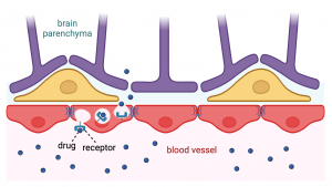
Laitinen’s PhD project is part of Obesity Curiosity and her goal in the first stage, is to develop a human arcuate nucleus model. Then in the second stage, they will use the model to study the effect of factors of interest, such as medications with weight gain as a side effect, on the human brain in vitro.

Currently, Laitinen is completing her research at a scientific collaboration visit in Dr Florian Merkle’s lab at the Institute of Metabolic Science University of Cambridge, funded by the Helsinki Institute of Life Science (HiLIFE) Undergraduate Student Research Trainee Scholarship. “My work here is focused on directed differentiation of human induced pluripotent stem cells (hiPSC) to hypothalamic neural origin cells, including neurons. This way we can generate a model that quite faithfully replicates human physiology without having to deal with the inaccessibility of the human brain otherwise. The hiPSC cells have been generated from volunteer donations of patients in past”, she explains.

“Working in Dr Merkle’s lab allows me to learn their robust methodology of hiPSC-derived 2D hypothalamic cell culture and with his generous support I am highly grateful of, I have expanded into preliminary testing of my own protocol to generate organoids. I hope to be able to establish corresponding cultures when I return to Helsinki. The research methods learnt during the exchange will be the firm foundation of my PhD research project, and forming strong collaboration will be immensely helpful. I am highly enthusiastic about this opportunity!”

Laitinen independently designed her new research opening involving a novel methodology of assembloids based on the literature of the field in 2022 and convinced Dr Aitta-Aho to become her supervising Principal Investigator in October of the same year. She has since executed her research in the Loss of Control Behaviour lab of Dr Aitta-Aho alongside her medical school studies at the University of Helsinki. Obesity Curiosity, the wider project involving multiple approaches to study the root causes of the obesity pandemic is their joint effort.

“To understand obesity, I have started the same way as many scientists before me, by finding special cases and trying to understand the general rule from there. Common clinical knowledge and scientifically proven fact is that many psychiatric drugs statistically cause patients to gain substantial amounts of weight in a relatively short period of time. I want to study this with a novel human brain model. Long-term, I hope to be able to help in improving the treatment and prevention of obesity

Before founding Obesity Curiosity, Laitinen had close to nine months of full-time biomedical research experience. After winning Millenium Youth Prize 2018 , she worked at Aalto University in the Department of Neuroscience and biomedical engineering rotating between different laboratories in summer 2019, exploring what the work of a researcher is like. Summer of 2020 and the four months between final exams and starting medical school in 2021, she worked at Professor Pekka Katajisto’s lab (HiLIFE Institute, University of Helsinki) in stem cell research.

In Katajisto lab, Laitinen investigated basal mammary gland cell adhesions to EMC proteins in the normal development and the cancer of mammary gland epithelium.

“The experience was amazing! Professor Pekka Katajisto and Dr Johanna Englund, my supervising researcher in the lab, always believed the most and best of me, teaching me to reach my full potential and ask all my questions. They also demonstrated how great supervising at best can be!”

Her work in 2021 also resulted in her first two co-authored publications, an accomplishment she is proud of and grateful for. “In the scientific world, they are proof that I am capable of executing research at the meticulous level, and maybe help to get respected as a fellow researcher regardless of my young age”

In her first year of medical school, she got accepted to the MD-PhD-program at the University of Helsinki, which had been her goal ever since she learnt that it is possible to become physician-doctor. “It was a nerve-wracking process, as one can apply for the program only once [in the first year of medical school] and I had focused my work towards that goal for a long time. Acceptance felt like reaching the top of a mountain – a completion – and a wonderful landscape of the research world opening in front of me!”

Her initial interest in cancer led to her choosing the first rotation lab studying ovarian cancer. She completed 2 months rotation in the Dr Anniina Färkkilä’s lab (Systems Oncology Research Program (ONCOSYS), University of Helsinki) in the summer of 2022 to learn about three cutting-edge methodologies: cyclic immunofluorescence method, patient-donated sample-derived organoids and complex cell typing flow cytometry.“The point of rotation is to get experience that helps with PhD projects, and I strongly believe these methods will help me design my own project and readouts to investigate my research questions”

“The burning question keeping me up at night is What causes the obesity pandemic? Ultimately this is about the health of over 700 million people suffering from obesity already today and countless others to come. Obesity affects everyone, if not directly themselves, their loved ones. I have a hypothesis, so I am trying my best to test it. I am working in molecular neuroscience to hopefully make the world a better place.”

Laitinen was awarded HiLIFE Undergraduate Student Research Trainee Scholarship for international research exchange in November 2022. She secured a position in Dr Merkle’s lab to conduct the research visit between May-August 2023 working on her Obesity Curiosity project.

Granting a Drug an ID Badge – Harnessing Receptor-Mediated Transcytosis for Crossing the Blood Brain Barrier

Imagine an agency headquarters with tight security policies in place. There are security guards on all doors and no entry without an ID badge. Various employees pass by the gate, all with different job titles and responsibilities. Entrance is also granted for the supportive personnel such as maintenance, cleaning services, and catering. Everybody knows their own specialized role and together with a strong management it is made sure that everything flows in a highly organized manner. Occasionally, an intruder tries to invade the establishment, but they are swiftly stopped by the security and police officers who arrive just a moment later to make sure nothing was stolen, and no one harmed. And if something or someone was to be injured, a team of healthcare professionals would arrive to take care of the situation.

In a similar way, in our bodies, molecules are travelling in the bloodstream and when wishing to enter the brain they arrive to the gate of the blood-brain barrier, or shorter the BBB. During my HiLIFE Research Trainee internship, I got an opportunity to join the Brain Targeting Program at the Wyss Institute for Biologically Inspired Engineering at Harvard University. Research in this translational program centers around the study of the BBB.

What is the Blood-Brain Barrier?

The BBB is a highly specialized structure consisting of endothelial cells that form the blood vessel wall or lumen. These cells are located very close to one another as they form the so-called tight and adherens junctions by binding to multiple proteins. This compact structure allows a limited number of molecules to pass through the BBB. Molecules can pass through via diffusion only if they are small enough or lipid soluble. Another mechanism to cross the BBB is by utilizing transport molecules which carry some substances from the blood to the brain parenchyma (that is, the functional tissue of the brain). Thus, only if the molecule has a correct ID badge, it can enter to the brain.

In the BBB, on the brain side, endothelial cells are enclosed by other cell types: pericytes and the end-feet of astrocytes that support the integrity of the barrier – similar to those security guards in the agency headquarters. To protect the brain from the occasional invasion of intruders, there are microglial cells which work with astrocytes to engulf unwanted molecules or injured cells, and produce cytokines which amplify inflammatory signals to increase the body’s response to the intruder. There is also an efflux mechanism that can expel the uninvited guests who manage to slip through the BBB. Not only do astrocytes contribute to the integrity of the BBB, but they also connect neurons to the vasculature. Thus, there is dynamic communication between the blood vessels and the brain to keep the entire structure highly functional – in our agency example they function like a network of guards with their security earpieces.

Together, the BBB with all the different cell types form the neurovascular unit. This specialized structure is distinct from any other blood vessels in the body, and it therefore also functions very uniquely. It maintains the regulation of blood, oxygen and nutrient flow, immune response, and waste clearance which are all crucial for the homeostasis – the self-regulated normal functioning – of the brain and the body.

Image representing blood vessel surrounded by pericyte and astrocyte cells. On the brain parenchyma side neurons and microglia.
Schematic view of the blood-brain barrier. Created with BioRender.com.

The Challenge for Delivery of Therapeutics into the Brain

As much as the BBB protects the brain from unwanted guests such as microbes and toxins, it also very effectively inhibits various drug molecules from reaching the brain. The entry of up to 98% of small-molecule drugs and 100% of larger macromolecular therapeutics is blocked by the BBB. This makes it difficult to treat different diseases affecting the central nervous system, such as neurodevelopmental and neurodegenerative diseases or brain tumors.

Various methods for enhancing brain drug delivery have been developed over decades, yet only a few have proven effective. Some of these technologies need surgery or other invasive methods such as deep brain stimulation, while others such as receptor-mediated, nanoparticle carrier, or focused ultrasound strategies are non-invasive. Our Brain Targeting Team at the Wyss Institute focuses on target discovery and validation utilizing the non-invasive receptor-mediated method, named as receptor-mediated transcytosis, for more efficient brain drug delivery. My main tasks involve handling human brain tissue samples and analyzing both transcriptomic and proteomic data to identify and assess the transport potential of numerous targets.

Receptor-mediated transporters are endogenous proteins located at the BBB blood vessel wall that can have various functions in the body. For example, the most studied receptor-mediated transporter, the transferrin receptor, normally functions in iron uptake from the cell membrane inside the cell. Antibodies can be engineered to bind to these receptors in a way that also other molecules than only the endogenous ones can be internalized. Antibodies linked with desirable therapeutics and designed to utilize these transporter targets are often referred to as Trojan horses, as they are only allowed to cross the BBB when in disguise, subsequently exerting their therapeutic effects once inside the brain. Thus, transferrin receptors can recognize an antibody while inadvertently allowing the entire complex to be carried in the brain without the body’s surveillance system recognizing the foreign drug molecule.

Blood vessel on the bottom on the image, lined by endothelial cells. On top pericytes that are enclosed by astrocyte end-feet. Drug from the blood vessel is carried across the blood-brain barrier with a receptor protein.
Schematic view of the receptor-mediated transcytosis for drug delivery across the blood-brain barrier. Created with BioRender.com.

I prefer to imagine that the BBB and therapeutic compounds are on the same side of the battle. Thus, instead of a Trojan horse, this technology harnesses the capabilities of our body so that the BBB offers a helping hand in drug delivery and welcomes an additional aid to battle against protein aggregates found from neurodegenerative diseases or tumor cells in brain cancers when our body and its cells need assistance. Ultimately, it is about granting an ID badge for the drug, for the invited guest in the headquarters of our wondrous body.

A little bit about me:

I am a Master’s student in Translational Medicine at University of Helsinki, currently located in Boston, United States, to conduct a six-month-long internship at the Wyss Institute at Harvard University. In my M.Sc. studies I specialize in translational neuroscience and personalized medicine. My Master’s thesis looked into the molecular mechanisms behind Alzheimer’s disease and while in the midst of my experiments, I got enchanted about the BBB. Therefore, I am deeply thankful and excited to contribute to the Brain Targeting Program at the Wyss where innovations are translated into clinical practice. My next HiLIFE Trainee blog post will provide more insights into my internship experiences, so stay engaged for more!

Mareena Hyypiä

From A Nearby Ditch to Lab Bench: Exploring the Soil Viriome


Hello! My name is Erika Nordman, and I am a second-year student in the Bachelor program of Molecular Biosciences at the University of Helsinki. I am thrilled to be one of the 2023 HiLIFE Research trainees and during my internship, I am working on characterising a novel soil bacteriophage.

When I started my studies in University of Helsinki in autumn 2021, I quickly discovered my interest in the micro-world, particularly in bacteriophages. Bacteriophages are viruses utilising the bacterium’s machinery to replicate and spread. What initially caught my attention about bacteriophages was their funky appearance and as I delved deeper into the fascinating world of bacteriophages, I surprised myself how intriguing these alien-like creatures truly are!


I started my traineeship last April under the lead of Minna Poranen and Hanna Oksanen at the Viikki campus in University of Helsinki. The group is involved in numerous research projects focused on studying viruses, and I feel honoured to have my own project within their group. So far, my internship has been very exciting for me, as becoming a virologist and scientist is my ultimate career goal in the future. Being able to participate in a research project within my own field of interest is a valuable opportunity to get during the early stage of my studies.

Applying Skills from a Lab Course in Research Work

Last autumn, I enrolled in a Helsinki University course “Practical Exercises of Bacteriology and Virology”. During our lab course, we collected a soil sample from the Viikki campus, enriched the sample and performed plaque assay using Bacillus cereus as the host bacterium for our experiment. The phage isolate was grown and purified by rate-zonal gradient ultracentrifugation and morphology of the bacteriophage was observed through transmission electron microscopy (TEM) and negative staining, and we determined the sizes of the main virion proteins using SDS-PAGE.

Our virus was assigned the name BCIP-1 (short for B. cereus infecting phage). BCIP-1 was found to have an icosahedral  structure and possibly contain a lipid-layer inside the capsid head. In the image below are our transmission electron microscopy pictures of BCIP-1 virions obtained from our laboratory course. Unfortunately, the virus capsid appears empty, suggesting that something in our purification protocol used during our lab course caused the loss of BCIP-1 genome and potential fragmentation of the virus’ head and its possible tail.

Since the main objective of the lab course was to learn the basic protocols in virology, there was limited time for troubleshooting and repeating the experiments. My traineeship goal is to optimise the conditions for successful virus purification, redo the transmission electron microscopy and conduct additional experiments to uncover other properties of BCIP-1. For example, I would like to verify the presence of the lipid layer inside the virion, determine whether the BCIP-1 genome consists of DNA or RNA and if the genome is circulated or linear.

Negative-stained transmission electron microscopy pictures of the icosahedral BCIP-1 virions from University of Helsinki lab course “Practical Exercises of Bacteriology and Virology” in autumn 2022.

The Power of Laboratory Courses in Virology Discoveries

A similar laboratory course was enrolled at the University of Jyväskylä in 2010, where a bacteriophage isolation from a boreal lake water sample led to the characterization of a new type of virus in the family Finnlakeviridae. The described virus is unique as it is currently the only known icosahedral internal membrane-containing virus containing a single-stranded DNA genome (Laanto et a., 2017).

The B. cereus bacteriophage that I have isolated has similar properties with the Finnlakeviridae representative virus called FliP (Flavobacterium-infecting phage). These characteristics include isolation from a boreal environment, icosahedral capsid morphology, and an inner lipid membrane. However, this virus infects a gram-positive bacterium while the host of FLiP is gram-negative. The similarities make my virus extremely intriguing, since they suggest a possibility that BCIP-1 could be evolutionarily close to FliP.


Light scattering zones indicate the migration of the virus in a sucrose gradient during rate-zonal virus purification. These zones are documented and collected, and the protein concentration of the purified virus is determined using the Bradford assay.

Unravelling the mystery:  Piece by Piece

So far, my journey with this virus has been filled with trials and errors. Since this bacteriophage has never been studied before, information is gathered bit by bit, combining and comparing various factors that could influence its ability to infect its host.

Currently I am concentrating on establishing buffering conditions to preserve the virions’ integrity and infectivity during virus purification. Once successful, I will have the opportunity to redo transmission electron microscopy and hopefully observe some intact virus particles! Ultimately, by comparing the characteristics identified with FLiP, my aim is to assess the potential of BCIP-1 being evolutionarily close to it and determine the possibility that BCIP-1 could belong to the same viral family.

Making novel viral discoveries is crucial, due to the immense number of viruses present in the environment, with only a fraction having been studied thus far. Viruses exhibit a vast genetic diversity within the soil, influencing the ecological dynamics in their respective ecosystems. Bacteriophages play a significant role in the horizontal gene transfer among their host organisms, maintaining the microbial homeostasis and contributing to nutrient circulation and organic matter decomposition. (Batinovic et al. 2019) By studying new viruses, we can enhance the understanding of the dynamics between viruses and their hosts, unraveling the mechanisms of viral infection, replication, and spread. Bacteriophages have wide-ranging applications in research and many biotechnological methods, and making novel viral discoveries fuels advancements in biotechnology, medicine, and bioremediation.

Everything Big Starts with Something Little

It has already been an extraordinary experience at this stage of my studies to witness how scientific projects can emerge from seemingly insignificant beginnings, such as collecting a spoonful of soil from a nearby ditch for a laboratory course. Step by step the puzzle of this bacteriophage is unraveled, and I have the privilege of being the first one to make discoveries about this virus. I am very curious to witness the pieces slowly come together enabling me a deeper understanding of this virus. Who knows, perhaps this virus is truly one-of-a-kind. I am looking forward to providing you with updates on my project in my upcoming HiLIFE blog post!

References:

Batinovic S, Wassef F, Knowler S, Rice D, Stanton C, Rose J, Tucci J, Nittami T, Vinh A, Drummond G, Sobey C, Chan HT, Seviour R, Petrovski S, & Franks A. 2019. Bacteriophages in Natural and Artificial Environments. Pathogens, 8(3), 100. https://doi.org/10.3390/pathogens8030100

Laanto E, Mäntynen S, De Colibus L, Marjakangas J, Gillum A, Stuart DI, Ravantti JJ, Huiskonen JT, Sundberg LR. 2017. Virus found in a boreal lake links ssDNA and dsDNA viruses. Proc Natl Acad Sci U S A. 114(31):8378-8383

My battle against RNases


Hi! I’m Anna and I’m a Master’s student at University of Helsinki (UH) in the programme of Genetics and Molecular Biosciences. I am honored to have been chosen as one of the HiLIFE research trainees for the year 2023.

For as long as I can remember I have been interested in life sciences. Nowadays, my main academic interests lie in functional genetics, RNA biology, translational biology, and biomedicine. I received my Bachelor’s degree at UH and wrote my thesis on the canonical and non-canonical functions of aminoacyl-tRNA synthetases. That was when my passion for RNA and translation really began. Learning about and understanding the complex systems that facilitate the basics for life is something that fascinates me deeply.In my spare time I am an active board member in the student organization Svenska Naturvetarklubben (SvNK), where I work as the vice chairman for the year 2023. A large portion of my spare time goes to the student organization, but I also love to draw and do some science related arts and crafts (such as making earrings out of the elegant tRNA secondary structure). 

RNA is part of the central dogma of molecular biology, but it has many other functions in the cell beyond that. There is a lot of ongoing research on a variety of different RNA types, but there is still a lot to learn about one of the first discovered, and no less fascinating, RNAs: tRNAs. For a long time, tRNAs had been thought to be mere vessels for amino acids, that their only job was to bring the amino acid to the ribosome for translation. tRNAs are indeed essential players in the basics of the translational system, but they also have many interesting regulatory functions. tRNAs have been shown to have a part in both transcriptional and translational regulation, as well as apoptotic pathways (Avcilar-Kucukgoze, I. & Kashina, A. 2020). Recently discovered tRNA derived RNA fragments have been shown to regulate both transcription and translation in miRNA-like ways and are seen as a part of the ncRNA family (Liu et al, 2022).  

The research in Docent Peter Sarin’s RNAcious laboratory focuses on molecular modifications on tRNA nucleotides. All natural tRNAs have molecular modifications on some nucleotides, such as methylation, acetylation and pseudouridylation. These modifications are important for the stability of the tRNA molecule, stress response in the cell, and regulation of translation (Koh & Sarin, 2018). For instance, hypomodification of the anticodon loop of tRNA molecules can disrupt anticodon recognition and may cause issues in protein homeostasis (Koh & Sarin, 2018). Diseases that have been associated with issues in the tRNA modificome, sometimes called tRNA modopathies, are e.g., microcephaly, intellectual disabilities, and multiple cancers (Chujo &Tomizawa, 2021). Modopathies can be caused by mutations or dysregulation of certain enzymes, such as methyltransferases and pseudouridine synthases, that regulate the modification profile of tRNAs (Chujo & Tomizawa, 2021).   

We still have a lot to learn about the role of tRNA modifications in health and in disease. As Chujo & Tomizawa (2021) mention, we do not have a complete understanding of the general cytoplasmic tRNA modification profile in healthy humans, which in turn makes it difficult to study modopathies since we have no healthy reference. A big obstacle in the research of tRNA modifications has been the loss of molecular modifications during sample handling as well as lack of methods to identify all modifications and their exact location. The RNAcious laboratory does e.g., research on method development to identify different types of modifications and research on the role of tRNAs and tRNA modifications in viral infection.    


The research in RNAcious. From RNAcious laboratory website, 21.6.2023, https://www.helsinki.fi/en/researchgroups/rnacious-laboratory/research-topics-0


My HiLIFE traineeship at RNAcious started in the beginning of April, and it has been a great pleasure to work in a prestigious, diverse, and international research group. So far, I am involved in a project where the aim is to characterize tRNA modifications in different mouse organs. In addition, I am working on a project where the goal is to plan, produce and isolate hypomodified tRNAs. My work has e.g., comprised of protocol optimization, RNA and tRNA isolation, mass spectrometry sample preparation, In vitro transcription (IVT), IVT template design and protein production and isolation. It has been a privilege to work with pioneers in the field and share and ponder different ideas with them.    


One thing that I have concluded thus far is that all RNA researchers have one common enemy, RNases. Ribonucleases: the enzymes that seek to destroy what is dearest to us, and they reveal their destruction through smeary bands in polyacrylamide gels. That is why I comprised a short list below which includes a few different ways you can inhibit RNase activity.  

RNA work 101:   

  • Use nuclease free eppendorfs.    
  • Use 3% Hydrogen peroxide to clean counter tops and gloves (anything that may be in contact with the sample).    
  • Always use gloves when working with RNA to not contaminate your sample with RNases from your skin.    
  • RNase inhibitors are your best friend.  
  • Always keep your sample on ice. 
  • Heating samples at 80°C for 5 min should inhibit RNase activity.  
  • Store your RNA in -80°C to inhibit degradation.   

I am excited to continue this RNAcious journey and fight my own battle against RNases!           


References: 

Avcilar-Kucukgoze, I. & Kashina, A. (2020). Hijacking tRNAs From Translation: Regulatory Functions of tRNAs in Mammalian Cell Physiology. Front Mol Biosci 7, 610617.

Chujo, T., & Tomizawa, K. (2021). Human transfer RNA modopathies: diseases caused by aberrations in transfer RNA modifications. The FEBS journal, 288(24), 7096–7122. 

Koh, C. S., & Sarin, L. P. (2018). Transfer RNA modification and infection – Implications for pathogenicity and host responses. Biochimica et biophysica acta. Gene regulatory mechanisms, 1861(4), 419–432.

Liu, B., Cao, J., Wang, X. Guo, C.,Liu, Y., Wang, T (2022). Deciphering the tRNA-derived small RNAs: origin, development, and future. Cell Death Dis13, 24. 

On Thin Ice: Baltic Ringed Seals in Peril


Have you ever seen a seal in Helsinki? Count yourself lucky if you have (I, for one, have yet to see one). However, there was a time when ringed and grey seals thrived throughout the Baltic Sea. They would gracefully search for fish along the coast and take leisurely breaks on beaches and sea ice. Sadly, today they have become a rare sight, with ringed seals in particular confined to a few scattered locations across the Baltic.

Now, here’s a little about me: I’m no biologist. In fact, my last biology course was almost a decade ago. However, those who knew me as a child can vouch for my infamous obsession with wildlife. After obtaining my Bachelor’s degree in engineering, I spent several years working in financial services. But now, after a decade-long detour, I find myself returning full circle to my childhood passion, albeit with a twist. Recently, I completed my first year in the Master’s Programme in Life Science Informatics (LSI) at the University of Helsinki, with a specialization in mathematical ecology.

Just a few weeks ago, I embarked on an exciting 4-month internship funded by HiLIFE. During this internship, I will be working alongside the Environmental and Ecological Statistics Group at the University of Helsinki and the Natural Resources Institute Finland (Luke). Together, we aim to develop mathematical and statistical models that can predict future population sizes of Baltic ringed seals, and if things go well, perhaps grey seals too. We are particularly interested in the effects of hunting, fishing and climate change.

A ringed seal (Pusa hispida).
Image credit: Kingfisher, CC BY-SA 3.0, via Wikimedia Commons

The Past and Present of Ringed Seals

The history of Baltic ringed seals stretches back over 10,000 years. As the glaciers receded during the last ice age, these remarkable creatures migrated into the Baltic region. Over time, the changing climate forced them further north, eventually leading to the isolation of the Baltic population from their Arctic brothers and sisters. Today, these seals are adapted to the unique conditions of the Baltic Sea, and are considered a distinct subspecies.


Unfortunately, the journey for Baltic ringed seals has not been without challenges. Humans have been hunting these seals since their arrival in the Baltic, initially employing traps, harpoons and nets, and later transitioning to firearms. In the early 1900s, conflicts with fisheries prompted the implementation of bounties on ringed seals in the Baltic states. These hunting practices, coupled with chemical contamination in the Baltic, posed severe threats to the population.

The consequences of these detrimental factors became evident as the population plummeted from an estimated 200,000 individuals in 1900 to just a few thousand by the 1970s. The chemical contamination led to widespread female sterility, exacerbating the decline. Today, the Baltic ringed seal population is fragmented into four sub-populations in the Bothnian Bay, the Gulf of Riga, the Gulf of Finland, and the Archipelago Sea.

Subpopulations of the Baltic ringed seal (Pusa hispida botnica) and their estimated population sizes.
Image adapted from: Halkka, Antti, and Petteri Tolvanen. “THE BALTIC RINGED SEAL.”

Future Threats

Despite some progress in mitigating the threats faced by Baltic ringed seals, uncertainties loom on the horizon. The ban on seal hunting and the discontinuation of using harmful substances such as DDTs and PCBs in the late 20th century have contributed to the recovery of ringed seal populations, particularly in the Bothnian Bay. However, the status of the remaining sub-populations remains uncertain.

In recent years, rising seal populations in the Bothnian Bay have led to conflicts with coastal fisheries. To address these conflicts, hunting has been reinstated as a management strategy. Balancing the needs of both seals and fisheries is a complex challenge that requires careful consideration and effective management decisions.

In addition to hunting and chemical contamination, Baltic ringed seals are threatened by entanglement in fishing nets. The unintentional capture of seals in fishing gear poses a potentially serious danger to their survival and calls for the development of sustainable fishing practices that minimize bycatch.

Another pressing concern that looms over Baltic ringed seals is climate change. These seals heavily rely on sea ice for their survival. They construct lairs on thick and stable sea ice, where they overwinter, give birth, and raise their pups. Sea ice also serves as a crucial resting and molting platform for them. However, as climate change accelerates, the loss of sea ice becomes an imminent threat to their habitat.

Illustration of a ringed seal pupping lair. The lair provides shelter to
newborn pups from harsh winter weather, predators and even pathogens. Loss of sea ice and reduced snowfall due to climate change are likely to have significant negative effects on pup survival.
Image credit: Robert Barnes, UNEP/GRID-Arendal.

The extent to which the loss of sea ice will impact seal populations remains uncertain. Predicting the future dynamics of seal populations in the face of climate change requires sophisticated mathematical and statistical models that can account for various ecological variables and complex interactions.

The Role of Mathematics and Statistics

In the realm of wildlife conservation and population dynamics, the use of mathematical and statistical models plays a crucial role. These models enable researchers to predict and understand the consequences of various management decisions, thereby aiding decision-making processes.

During my HiLIFE funded research project with the Environmental and Ecological Statistics Group, my goal is to construct a Bayesian State Space Model (SSM), which is a type of hidden process model. As the name suggests, hidden process models aim to infer processes that cannot be directly observed. In the case of Baltic ringed seals, our knowledge of the population is based on hunting reports, interviews with fisherman, and annual surveys that count the seals hauled out on ice. However, the true underlying process, encompassing the births, lives and deaths of seals, remains hidden from our direct observation.

Bayesian SSMs provide a powerful tool to unravel the hidden dynamics of seal populations. By combining available data with probabilistic modeling techniques, we can make informed inferences about what is happening “behind the curtain”. These models enable us to estimate demographic rates, assess population trends, predict the effects of management decisions, and gain deeper insights into the complex dynamics of Baltic ringed seals.

A candid photo of my deskmates.

My First Weeks on the Job

During my initial days, I dedicated a significant amount of time reading up on ringed seal biology and learning about the application of state-space models (SSMs) in wildlife population dynamics. Once I gained a reasonable understanding of the ringed seal lifecycle, I constructed a simple age-structured model of their population dynamics. Through the use of Bayesian techniques, I inferred key vital rates, such as age-dependent fertility and mortality rates, achieving a good fit to the available data.

Over the next few months, my objective is to gradually enhance the complexity of this model, striving to develop an SSM that incorporates the intricate mechanistic details of ringed seal biology, as well as the effects of hunting, fishing and the loss of sea ice due to climate change. Among the major challenges ahead is the modeling and inference of density-dependent processes. No population can grow perpetually. Whether it’s the availability of food, space, or the presence of predators, there will inevitably be limiting factors. Unraveling these factors for ringed seals poses a significant challenge, especially since the population is currently rebounding from historically low numbers. However, understanding these limitations is crucial if we are to make meaningful predictions about the future of ringed seals.

It is precisely these kinds of challenges that make research truly exhilarating, and I consider myself fortunate to be confronted with them!

On how colorful fish fall in love in the dark

Have you ever stared closely at fish in an aquarium? Isn’t it just amazing to look at the plethora of shapes, colors, and different behaviors that the fish, living on the other side of the glass, display? My younger self really enjoyed the wonders of the aquarium hobby – I believe the underwater world bears some of the world’s most amazing representations of life, and those tanks could, in some way (ethical issues aside), depict a little part of these marvels. But, how do fish see each other? Are they attracted to the colors other fish display? If so, what happens then, in the dark?

I am Adrián Colino Barea, a Spanish wildlife biologist, and a current first year student of the Master’s Programme in Ecology and Evolutionary Biology at the University of Helsinki. My main motivation to join this programme here was to dig deeper in applied ecology in tropical regions, as the University of Helsinki has several research lines in the tropics. I am aiming to focus on biodiversity conservation: I want to contribute and make a difference to revert the trends that current human-driven impacts have on nature globally.

Not surprisingly, me in the outdoors, hiking, looking for some interesting endemics in Azores. Greatly attracted by field ecology, I would not have pictured myself doing this exciting lab-based internship back when this photo was taken!

While my passion has always been working on the field, touring the fish room of the Integrative Evolutionary Biology (IntEvoBio) lab during an introductory course of the master’s last year awakened my younger self thrill. Racks with tens of tanks full of cichlid fish from the African Great Lakes fill the room, all around us, while some of the IntEvoBio lab members, led by Prof. Claudius Kratochwil, guided us through the nuts and bolts of their interesting job with all these fish, and why they are so particularly wonderful.

African cichlids: the book example of speciation

The African Great Lakes are three massive freshwater bodies in tropical West Africa, originated by ancient tectonic shifts. However, these lakes have borne livable conditions for a relatively short time. A certain group of cichlid fish got to these waters and started evolving at an unprecedented rate, fitting every possible niche, and generating entire, complex ecosystems, all based in different cichlids. What is more, they display very different color patterns of colors, bands, and spots, and perform elaborated mating rituals. Their unusually fast evolutionary process is based in ‘sexual selection’, meaning that individuals choose others as partners based on certain traits of their preference, despite the fact that these traits could make them, for instance, more showy – and therefore easier to spot by predators, hence against ‘natural selection’ as described by Charles Darwin.

To put it in perspective, for any evolutionary biologist, the cichlids of the African Great Lakes are old acquaintances, and for some they are the entire reason for their research. For many biologists, they are just inexplicably special. Some even say that the basis of evolution today would be radically different, had Darwin visited these lakes instead of the Galápagos Islands almost 200 years ago, and looked to these fish.

While in the lab, I enjoyed seeing all of the complexity that I have read about, right before my eyes. The researchers explained to us how these fish (and their evolutionary process) strongly rely on color communication. They have developed color signaling to inform about their sexual status, the hierarchy within a group, and even some species can change their colors rapidly, to give fast information! 

Cichlids have evolved into a big variety of colors, shapes, and behaviors among the waters of the three African Great Lakes. Just to clarify, the big guy on top left is not a cichlid 😉

However, one of the drawbacks to this very fast evolutionary process is that it is also very fragile, as species may seem radically different in colors and shapes but have very similar DNA, because of how recently they have evolved. In addition to this potentially fragile evolution, these lakes are particularly vulnerable to the impacts of human development. Lake Victoria is the second-largest lake on the planet by area and a huge population increase, has led to 40 million people now inhabiting the coastal region This human activity has resulted in major issues related to biodiversity, mostly through pollution, overfishing, and introduction of invasive predator fish and floating plants. 

How does sexual selection work under deterred underwater light conditions?

In particular, an invasive floating plant, the water hyacinth (Pontederia crassipes), is causing terrible damage to all ecosystems in Lake Victoria, and migh spread to other lakes. These plants prevent light from penetrating, hindering the oxygen production in the water and making the ecosystems within it collapse. Moreover, the human communities around the lake cannot physically access their resources because of how dense and inexpugnable the floating mats are.

But, coming back to my initial concern, floating plants – as well as increases in turbidity – make the underwater dark. Is there anything happening directly to the cichlids? If they rely so strongly on visual communication for their daily lives and long-term evolutionary process, how do they cope under a scenario in which their vision – light supply – is hindered? How can they find and recognize each other effectively and ‘sexually’ select their preferred partners according to visual signals (colors)?

During my internship, beyond my research project, I am learning the basics of tank care and manipulating fish. I certainly work with beautiful animals, such as this Pseudotropheus (Chindongo) demasoni, and the yellow females of Ps. (Ch.) saulosi in the bucket.

Back during my lab visit, I asked some researchers and they didn’t have an answer. This visit was inspiring enough to raise meaningful biological questions, with profound biodiversity conservation meanings. I am very pleased that HiLIFE gave me the chance to shed light on this topic, by granting me a Research Trainee Scholarship. Since March, I can call Prof. Claudius Kratochwil my supervisor, and some of the PhD students that once showed me through their research, my helpful mentors. 

From March to today: the beginning of my experimental phase

During these months, I have been setting up my experimental arena, and dedicatedly come up with a protocol to follow. I am working on the mate choice of cichlid males over females under different light conditions. Only last week I started the experimental phase, and I am excited to know what will happen with the results that I am obtaining – I will keep you posted on my progress. If this experiment arise meaningful outcomes, these invasive floating plants would potentially be given a new threat to nature, and hopefully a new reason to focus efforts and find an effective solution in the field, for the cichlids, for the people in the region, and for the entire ecosystems.