Fireballs of ischemic stroke

Hi Everyone!

My name is Katariina Järvinen, and I am a second-year master’s student in the Translational Medicine program at the University of Helsinki. I conducted my master’s thesis research during summer 2020. One of the aims of my project was to evaluate the potential of different phagocytosis related genes on enhancement of microglia phagocytosis.

Working as a HiLIFE research trainee in Mikko Airavaara’s research group, I learned many new skills, for example culturing of cells, and gained valuable practical experience from working in a laboratory. In addition, I was able to widen my professional network. Overall, I was impressed with how the whole group works together: if something doesn’t go as planned, there is always help available and multiple people giving ideas on tackling the issue or guidance in proceeding with another suitable method.

Here is a picture of BV2 microglia 1 hour after adding phagocytosis bioparticles. Inside the cells the bioparticles react to pH change and give red fluorescence signal.

Being HiLIFE Research Trainee met all of my expectations despite COVID-19 restrictions. I’m happy that I got to do my traineeship in a research group that is doing research that excites me and that my project has high scientifical value to the members of the group. The results gained during the traineeship will be validated in ischemic stroke in vivo study.

I am deeply grateful to Mikko Airavaara and my supervisor Helike Lõhelaid for their support and guidance during my traineeship and after it. I would also like to thank HiLIFE for supporting me in my early career in medical research.

Here you can read more about neuroprotection and neurorepair group.

Katariina Järvinen

Leiomyomas, bioinformatics, a little bit of lab work and wonderful people

Circos plot

Hi, all of you reading this HiLife trainee blog! I am Vilja and I am a master’s student in genetics and molecular biosciences. At the beginning of this year, I was looking for a master’s thesis position, and I wished to find a project related to cancer or other tumors. I also wanted to further develop my skills on bioinformatics. I was super excited to get a master’s thesis position at Pia Vahteristo’s research group studying gynecological tumor genomics. I was supposed to start with my master’s thesis project in June, but the COVID-19 pandemic changed my plans. In the middle of August, I was happy to finally start with a very interesting project.

In my master’s thesis project, I am analyzing the genomic data of uterine leiomyomas. Uterine leiomyomas are benign smooth muscle tumors of the uterus. The prevalence is up to 70–80% in women. I am focusing on structural variants, such as translocations, inversions, insertions, huge deletions and amplifications. The HMGA2-RAD51B translocation is one of the best characterized structural variants in leiomyomas. This means that part of the chromosome 12 is attached to a part of the chromosome 14, so that HMGA2 receives active regulatory elements of RAD51B. This leads to a much higher expression of HMGA2 in tumor cells compared to normal cells. In addition to this well-known translocation, we have been lucky to come across and characterize some other structural variants in uterine leiomyomas.

Structural variants can be studied in many ways. Sometimes, if you have a hunch of the putative genes mutated and you are lucky enough, you might find something interesting by simply visualizing genomic data on IGV or some other visualizing software showing the alignment of paired-end data. There are also many bioinformatics tools which look for structural variants by utilizing algorithms. These tools, such as Delly, are analyzing discordant reads and split reads of the paired-end data. Also, bioinformatics tools analyzing read coverage can be used to detect copy number variations. Once you have found a structural variant, you may want to validate your finding. This can be done by using PCR and Sanger sequencing. These steps are exactly the ones that I am using for my master’s thesis project.

Circos plots are a nice tool for visualizing these structural variants. In the figure, you can see one of the preliminary Circos plots I have made by using RCircos (R package). Lines show translocations between chromosomes and intrachromosomal inversions, and the circular heatmap indicates copy number variations.

I have learned so many new things during the process, and the project is not even completed yet. For example, I have developed my skills in bioinformatics and learned about genetics and tumorigenesis of leiomyomas. I have also gained a better understanding of how research projects are performed and how technical difficulties can be approached and tackled. I am super lucky that I have had such great supervisors and colleagues, who I really want to thank. Also, I am very grateful for the support I have received from HiLife. It has been extremely valuable.

Vilja Jokinen

DNA, comets and shaky hands

Hi everyone!

My name is Piia Karhu and I am a first-year student in The Master’s Programme in Human Nutrition and Food-Related Behaviour at the University of Helsinki. I received a HiLIFE scholarship for the summer 2020 to work in a research project of my interest. I completed my research internship at the Viikki Molecular Nutrition group. The group studies the molecular mechanisms by which diets, foods and diet-derived compounds mediate their effects on health and prevention of non-communicable diseases. I was very happy to join this group as molecular nutrition is a very fascinating field of science. Here you can read more about Viikki Molecular Nutrition group.

During my summer, I was privileged to take part in the study that examines the impact of dietary habits on faecal water genotoxicity.  In the study, colonial epithelial cells were exposed to feacal water. Feacal water was extracted from stool samples collected during dietary intervention with healthy volunteers and the DNA damage created by the faecal water was examined. Purpose was to study whether there are differences in DNA damage of the cells when exposed to fecal water from volunteers that followed either plant protein diet, animal protein diet or diet that contains half plant protein and half animal protein. We measured the DNA damage using single-cell gel electrophoresis known as Comet assay. It is a commonly used and straightforward method for measuring DNA strand breaks in cells. The term “comet” refers to the leaked-out DNA of the cell as it often resembles a comet. The more intensive the comet tail is relative to the head of the cell, the more the cell is damaged.

microscope image of green cell comets
Here is an example picture of the comets

I had very interesting and mind opening summer job as I have never worked in this kind of project before. I acquired valuable experience working in the lab, especially in the cell lab and learned how to culture and handle cells. Working in a cell lab was a very nice experience even though it took a while before my hands stopped shaking! During my summer I gained a lot of practical experience, which will boost my confidence working as a researcher in the future.

I want to thank my supervisor Anne-Maria Pajari for this opportunity to work in her research group and Hana Slabá for excellent guidance during the summer. Also, I am deeply grateful to HiLIFE for supporting me, this was a very valuable experience!

This summer I didn’t sleep so well  

Sleeping wired

Paavo TeitinenHi everyone! My name is Paavo and I’m a neuroscience master’s student in University of Helsinki. In this blog text, I will share my experience working as a HiLIFE trainee in summer 2020.

First a little bit of background information. My previous academic background is in exercise physiology. However, even while studying sport sciences I was very interested in neuroscience, especially in the effect of sleep and stress on health. Therefore, I came to continue my studies in Helsinki as there are many excellent research groups here focusing on sleep. That is why I was (and I am) beyond excited, when I got a chance to do my HiLFE traineeship in the Sleep and Health -research group, led by professor Tiina Paunio.

Fun fact: in our first meeting I found out that we both share a background in track and field. I remember Tiina saying: “even though some work may not always be pleasant we can remind ourselves that it is nothing compared to 200 m intervals”. I must agree.

As I had no previous experience in sleep research, my traineeship started with learning polysomnography (PSG) and sleep scoring. PSG is the golden standard of the field, where one attaches a bunch of electrodes to the subject to measure their brain activity, muscle tone, heart rate and breathing. Sleep scoring means determining different macro- and microstructures of sleep from the (PSG) data. I was lucky to have Tuula Tanskanen as my scoring mentor. At first scoring was like being at optician when your glasses are not up to date: Tuula asked what this 30s epoch looks like, I squeezed my eyes looking at the signal, thought a bit and ended up half guessing. Luckily, practice trains your eye to find things from the signal, and I would like to think that the glasses I’m wearing now are at least close to the correct prescription. In addition to learning PSG and sleep scoring, I begun the basics of programming. Even though I’m still at beginner level, programming skills will for sure prove to be useful in further analyses of the sleep data.

charts
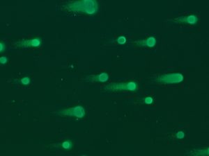
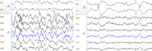
Here is an example of what different sleep stages look like. On the left there is slow wave sleep (N3) with large delta waves on the EEG channels. On the right there is REM sleep with the signature rapid eye movements on the top two EOG channels.

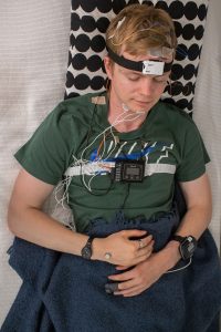
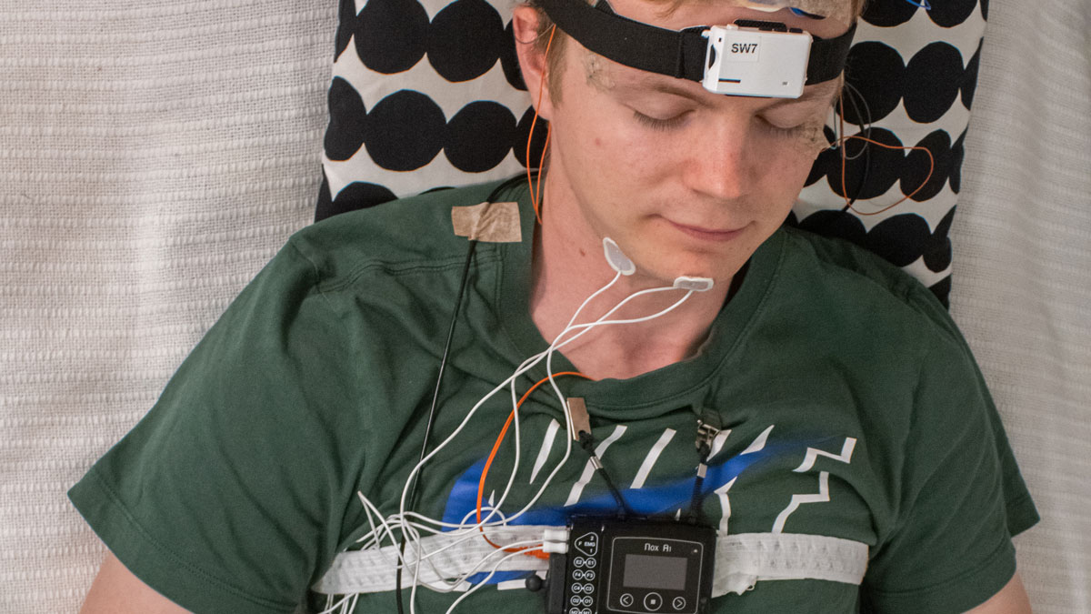
By this time, I’m sure all of you who read the title of the blog are shouting to the screen: “but Paavo why didn’t you sleep well?”. Well I’m glad you asked. You see, in addition to the thesis work I was doing, my job was to test and learn to use some new sleep monitoring devices. These devices will possibly be used in future projects and what better way to learn to use them than to wear them yourself! You can try to guess how many recording devices I’m wearing in the picture and what they are recording. The correct answer will be revealed at the end of this post.

Paavo wired up
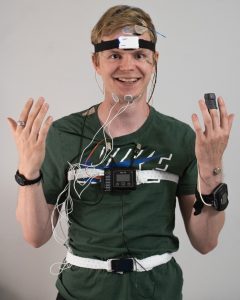
Would you participate in a sleep study if the researcher looked like this?

Based on the experience wearing these different devices, I found a direct correlation between the number of cables attached to my head and me being grumpy next morning. The relationship seems to be causal. However, the effect was strongly modulated by the successfulness of the measurement: couple of times I found that nothing was recorded, which was followed by a spike in blood pressure. Testing new equipment always comes with unexpected challenges. Solving these challenges included both reading theory to understand how the devices should work and trying things in practice to solve how they actually work. Couple of times I managed to combine the two: nothing worked, and I had no clue why. Jokes aside, I really enjoyed testing the devices and I could always ask help if needed.

I can honestly say that the HiLIFE traineeship has made a difference for my academic career. Even though I worked most of the time remotely due to covid-19, being able to participate in different (online) meetings and discussing things with my supervisor has opened many opportunities for the future. It is quite likely that I will continue to PhD studies in the same group. A word of advice for future trainees: don’t be afraid to ask and discuss things with members of the research group, you will be surprised where those conversations will lead you. Even the worst-case scenario is rarely worse than running 200 m intervals – and that was one of my favorite track sessions.

Special thanks for my sleep scoring mentor Tuula Tanskanen, for my ask me anything -person Tiina Härkönen and of course for my supervisor and the voice of reason Prof. Tiina Paunio. Thank you HiLIFE for enabling this amazing opportunity.

Paavo wired up and sleeping

  1. For those dying to know, the number of recording devices in the picture was five:

1) The black box on the chest is a polysomnography (PSG) device with several EEG, EOG and EMG electrodes, the white belt and pulse oximeter on the left hand (there are usually even more wring going with nasal airflow sensor and ECG electrodes that are not shown in the picture).

2) The white device on the forehead is an EEG headband, which measures brain activity, movement and temperature.

3) The watch-looking device on the right hand is an actigraphy monitor, which is an accelerometer measuring movement throughout the day.

4) The ring on the right-hand index finger measures heart rate, movement and temperature.

5) The small button on right hand (in the first picture on left hand – the deceit is revealed, the pictures were staged) is a tiny thermometer measuring changes in temperature.

Thinking about thoughts – my Cambridge Neuroscience experience

Text by Nejc Kejzar – HiLife trainee 2019

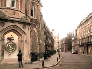
A sense of profound stillness settled upon me when I first set foot onto the cobbled streets of Cambridge. While gazing down Trinity Lane (pictured above), the scene momentarily took on a sephia tone as I was transported into the past. Some in animated conversation, others lost deep in their own thoughts, the scientific greats came streaming past me. There went James Clerk Maxwell, formulating the unification of electricity and magnetism in his mind. Niels Bohr arguing with Max Born over the best representation of quantum mechanics, timidly observed from the side by Paul Dirac, keeping his own to himself. Stephen Hawking playfully teasing the unphased Isaac Newton to try and reveal the gravitational secrets of black holes, followed by Alan Turing wondering just what makes their conversation distinguishable from machines. This surreal procession, stretching over hundreds of years and across disciplines, made me realize to what a special place I have arrived and that it is now my time – as Newton put it – “to see further by standing on the shoulders of Giants.”.

In Cambridge, emerald green grass is never far away from gothic architecture. This interwoven combination gives the whole town a studious feeling, which I used to my advantage on many a walk – there is nothing quite like retracing footsteps of great thinkers of the past and present to help solve a difficult problem of your own. Although fairly small in size, Cambridge contains many hidden alleys and secluded spots, creating a scholars’ paradise. What is more, the University permeates every street corner, lane and building. In fact, what makes Cambridge so special is that in a way, the town itself is the University. Departments, colleges and libraries from centuries past rub their brick shoulders with modern restaurants and bookstores. While one door might deliver to you a pint of London Stout shared with fellow intellectuals in the cosy athmosphere of one of 110 pubs, the neighbouring might open to reveal the interior of a physics’ lab – which, judging from an impressive number of novel discoveries streaming from Cambridge, is a rather harmonious relationship. It goes without saying, that the whole package has quite the Harry-Potteresque feel to it – after all, Hogwarts was inspired by dining halls, chapels and traditions of the University colleges. One of the formal dinners that I got to experience in Trinity College great dining hall (below) is a particularly fond memory – short of the floating candles and headless ghosts, I was hard-pressed to tell the difference from the Start-of-Term Feast at the School of Witchcraft and Wizardry. Regardless of whether you are a “Potterhead” or not, the subtle magical feeling is undeniable.


Whilst in Cambridge, I conducted my Master’s Thesis research in the Neurobiology Division of MRC Laboratory of Molecular Biology. Nestled among flowering fields (above), the imposing structure houses state-of-the-art laboratories and the highest concentration of experts in their respective fields that I have encountered to date (including 2 out of a total of 16 Nobel Laureates originating from this institute – casually saying “hi” to a passing Nobel Laureate on your way to morning coffee is quite an experience). The significance of the previous sentence with respect to the HiLIFE Trainee Scholarship cannot be overstated. Thanks to this scholarship, I not only got access to immense research resources, but could also closely interact with leading scientists. Being able to knock on a door instead of writing an email, awaiting a doubtful reply, makes a world of difference.             

My main focus here was studying the dynamics of AMPA receptors using molecular dynamics simulations. It is in part thanks to these receptors that we can learn and form memories. Therefore, in essence, I spent most of my time thinking about AMPA receptors using my AMPA receptors – quite an amusing thought. But the research I conducted in Cambridge is for me personally of greater importance still – it is through my research that I became involved in the Cambridge Neuroscience community and discovered my passion – merging the studies of artificial and biological intelligence. Prior to arriving at Cambridge I was not quite sure about where to take my career after graduating from Helsinki, but thanks to this experience, my future path is set and is looking as exciting as ever!

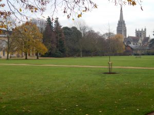
Alas, my 7 months in Cambridge have been gobbled up by the enigmatic Chronophage of the Corpus Christi clock. This has truly been an enriching experience and it is now up to you, my dear reader, to apply for HiLIFE Trainee Scholarships. For myself, the good-bye is only temporary, as I shall be returning to this enchating little town in October to start my PhD in Neuroscience, working on our brains’ internal GPS system!