My battle against RNases


Hi! I’m Anna and I’m a Master’s student at University of Helsinki (UH) in the programme of Genetics and Molecular Biosciences. I am honored to have been chosen as one of the HiLIFE research trainees for the year 2023.

For as long as I can remember I have been interested in life sciences. Nowadays, my main academic interests lie in functional genetics, RNA biology, translational biology, and biomedicine. I received my Bachelor’s degree at UH and wrote my thesis on the canonical and non-canonical functions of aminoacyl-tRNA synthetases. That was when my passion for RNA and translation really began. Learning about and understanding the complex systems that facilitate the basics for life is something that fascinates me deeply.In my spare time I am an active board member in the student organization Svenska Naturvetarklubben (SvNK), where I work as the vice chairman for the year 2023. A large portion of my spare time goes to the student organization, but I also love to draw and do some science related arts and crafts (such as making earrings out of the elegant tRNA secondary structure). 

RNA is part of the central dogma of molecular biology, but it has many other functions in the cell beyond that. There is a lot of ongoing research on a variety of different RNA types, but there is still a lot to learn about one of the first discovered, and no less fascinating, RNAs: tRNAs. For a long time, tRNAs had been thought to be mere vessels for amino acids, that their only job was to bring the amino acid to the ribosome for translation. tRNAs are indeed essential players in the basics of the translational system, but they also have many interesting regulatory functions. tRNAs have been shown to have a part in both transcriptional and translational regulation, as well as apoptotic pathways (Avcilar-Kucukgoze, I. & Kashina, A. 2020). Recently discovered tRNA derived RNA fragments have been shown to regulate both transcription and translation in miRNA-like ways and are seen as a part of the ncRNA family (Liu et al, 2022).  

The research in Docent Peter Sarin’s RNAcious laboratory focuses on molecular modifications on tRNA nucleotides. All natural tRNAs have molecular modifications on some nucleotides, such as methylation, acetylation and pseudouridylation. These modifications are important for the stability of the tRNA molecule, stress response in the cell, and regulation of translation (Koh & Sarin, 2018). For instance, hypomodification of the anticodon loop of tRNA molecules can disrupt anticodon recognition and may cause issues in protein homeostasis (Koh & Sarin, 2018). Diseases that have been associated with issues in the tRNA modificome, sometimes called tRNA modopathies, are e.g., microcephaly, intellectual disabilities, and multiple cancers (Chujo &Tomizawa, 2021). Modopathies can be caused by mutations or dysregulation of certain enzymes, such as methyltransferases and pseudouridine synthases, that regulate the modification profile of tRNAs (Chujo & Tomizawa, 2021).   

We still have a lot to learn about the role of tRNA modifications in health and in disease. As Chujo & Tomizawa (2021) mention, we do not have a complete understanding of the general cytoplasmic tRNA modification profile in healthy humans, which in turn makes it difficult to study modopathies since we have no healthy reference. A big obstacle in the research of tRNA modifications has been the loss of molecular modifications during sample handling as well as lack of methods to identify all modifications and their exact location. The RNAcious laboratory does e.g., research on method development to identify different types of modifications and research on the role of tRNAs and tRNA modifications in viral infection.    


The research in RNAcious. From RNAcious laboratory website, 21.6.2023, https://www.helsinki.fi/en/researchgroups/rnacious-laboratory/research-topics-0


My HiLIFE traineeship at RNAcious started in the beginning of April, and it has been a great pleasure to work in a prestigious, diverse, and international research group. So far, I am involved in a project where the aim is to characterize tRNA modifications in different mouse organs. In addition, I am working on a project where the goal is to plan, produce and isolate hypomodified tRNAs. My work has e.g., comprised of protocol optimization, RNA and tRNA isolation, mass spectrometry sample preparation, In vitro transcription (IVT), IVT template design and protein production and isolation. It has been a privilege to work with pioneers in the field and share and ponder different ideas with them.    


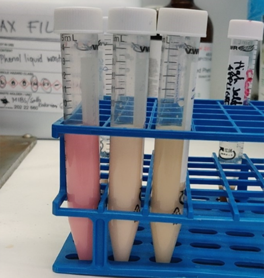
One thing that I have concluded thus far is that all RNA researchers have one common enemy, RNases. Ribonucleases: the enzymes that seek to destroy what is dearest to us, and they reveal their destruction through smeary bands in polyacrylamide gels. That is why I comprised a short list below which includes a few different ways you can inhibit RNase activity.  

RNA work 101:   

  • Use nuclease free eppendorfs.    
  • Use 3% Hydrogen peroxide to clean counter tops and gloves (anything that may be in contact with the sample).    
  • Always use gloves when working with RNA to not contaminate your sample with RNases from your skin.    
  • RNase inhibitors are your best friend.  
  • Always keep your sample on ice. 
  • Heating samples at 80°C for 5 min should inhibit RNase activity.  
  • Store your RNA in -80°C to inhibit degradation.   

I am excited to continue this RNAcious journey and fight my own battle against RNases!           


References: 

Avcilar-Kucukgoze, I. & Kashina, A. (2020). Hijacking tRNAs From Translation: Regulatory Functions of tRNAs in Mammalian Cell Physiology. Front Mol Biosci 7, 610617.

Chujo, T., & Tomizawa, K. (2021). Human transfer RNA modopathies: diseases caused by aberrations in transfer RNA modifications. The FEBS journal, 288(24), 7096–7122. 

Koh, C. S., & Sarin, L. P. (2018). Transfer RNA modification and infection – Implications for pathogenicity and host responses. Biochimica et biophysica acta. Gene regulatory mechanisms, 1861(4), 419–432.

Liu, B., Cao, J., Wang, X. Guo, C.,Liu, Y., Wang, T (2022). Deciphering the tRNA-derived small RNAs: origin, development, and future. Cell Death Dis13, 24. 

On Thin Ice: Baltic Ringed Seals in Peril


Have you ever seen a seal in Helsinki? Count yourself lucky if you have (I, for one, have yet to see one). However, there was a time when ringed and grey seals thrived throughout the Baltic Sea. They would gracefully search for fish along the coast and take leisurely breaks on beaches and sea ice. Sadly, today they have become a rare sight, with ringed seals in particular confined to a few scattered locations across the Baltic.

Now, here’s a little about me: I’m no biologist. In fact, my last biology course was almost a decade ago. However, those who knew me as a child can vouch for my infamous obsession with wildlife. After obtaining my Bachelor’s degree in engineering, I spent several years working in financial services. But now, after a decade-long detour, I find myself returning full circle to my childhood passion, albeit with a twist. Recently, I completed my first year in the Master’s Programme in Life Science Informatics (LSI) at the University of Helsinki, with a specialization in mathematical ecology.

Just a few weeks ago, I embarked on an exciting 4-month internship funded by HiLIFE. During this internship, I will be working alongside the Environmental and Ecological Statistics Group at the University of Helsinki and the Natural Resources Institute Finland (Luke). Together, we aim to develop mathematical and statistical models that can predict future population sizes of Baltic ringed seals, and if things go well, perhaps grey seals too. We are particularly interested in the effects of hunting, fishing and climate change.

A ringed seal (Pusa hispida).
Image credit: Kingfisher, CC BY-SA 3.0, via Wikimedia Commons

The Past and Present of Ringed Seals

The history of Baltic ringed seals stretches back over 10,000 years. As the glaciers receded during the last ice age, these remarkable creatures migrated into the Baltic region. Over time, the changing climate forced them further north, eventually leading to the isolation of the Baltic population from their Arctic brothers and sisters. Today, these seals are adapted to the unique conditions of the Baltic Sea, and are considered a distinct subspecies.


Unfortunately, the journey for Baltic ringed seals has not been without challenges. Humans have been hunting these seals since their arrival in the Baltic, initially employing traps, harpoons and nets, and later transitioning to firearms. In the early 1900s, conflicts with fisheries prompted the implementation of bounties on ringed seals in the Baltic states. These hunting practices, coupled with chemical contamination in the Baltic, posed severe threats to the population.

The consequences of these detrimental factors became evident as the population plummeted from an estimated 200,000 individuals in 1900 to just a few thousand by the 1970s. The chemical contamination led to widespread female sterility, exacerbating the decline. Today, the Baltic ringed seal population is fragmented into four sub-populations in the Bothnian Bay, the Gulf of Riga, the Gulf of Finland, and the Archipelago Sea.

Subpopulations of the Baltic ringed seal (Pusa hispida botnica) and their estimated population sizes.
Image adapted from: Halkka, Antti, and Petteri Tolvanen. “THE BALTIC RINGED SEAL.”

Future Threats

Despite some progress in mitigating the threats faced by Baltic ringed seals, uncertainties loom on the horizon. The ban on seal hunting and the discontinuation of using harmful substances such as DDTs and PCBs in the late 20th century have contributed to the recovery of ringed seal populations, particularly in the Bothnian Bay. However, the status of the remaining sub-populations remains uncertain.

In recent years, rising seal populations in the Bothnian Bay have led to conflicts with coastal fisheries. To address these conflicts, hunting has been reinstated as a management strategy. Balancing the needs of both seals and fisheries is a complex challenge that requires careful consideration and effective management decisions.

In addition to hunting and chemical contamination, Baltic ringed seals are threatened by entanglement in fishing nets. The unintentional capture of seals in fishing gear poses a potentially serious danger to their survival and calls for the development of sustainable fishing practices that minimize bycatch.

Another pressing concern that looms over Baltic ringed seals is climate change. These seals heavily rely on sea ice for their survival. They construct lairs on thick and stable sea ice, where they overwinter, give birth, and raise their pups. Sea ice also serves as a crucial resting and molting platform for them. However, as climate change accelerates, the loss of sea ice becomes an imminent threat to their habitat.

Illustration of a ringed seal pupping lair. The lair provides shelter to
newborn pups from harsh winter weather, predators and even pathogens. Loss of sea ice and reduced snowfall due to climate change are likely to have significant negative effects on pup survival.
Image credit: Robert Barnes, UNEP/GRID-Arendal.

The extent to which the loss of sea ice will impact seal populations remains uncertain. Predicting the future dynamics of seal populations in the face of climate change requires sophisticated mathematical and statistical models that can account for various ecological variables and complex interactions.

The Role of Mathematics and Statistics

In the realm of wildlife conservation and population dynamics, the use of mathematical and statistical models plays a crucial role. These models enable researchers to predict and understand the consequences of various management decisions, thereby aiding decision-making processes.

During my HiLIFE funded research project with the Environmental and Ecological Statistics Group, my goal is to construct a Bayesian State Space Model (SSM), which is a type of hidden process model. As the name suggests, hidden process models aim to infer processes that cannot be directly observed. In the case of Baltic ringed seals, our knowledge of the population is based on hunting reports, interviews with fisherman, and annual surveys that count the seals hauled out on ice. However, the true underlying process, encompassing the births, lives and deaths of seals, remains hidden from our direct observation.

Bayesian SSMs provide a powerful tool to unravel the hidden dynamics of seal populations. By combining available data with probabilistic modeling techniques, we can make informed inferences about what is happening “behind the curtain”. These models enable us to estimate demographic rates, assess population trends, predict the effects of management decisions, and gain deeper insights into the complex dynamics of Baltic ringed seals.

A candid photo of my deskmates.

My First Weeks on the Job

During my initial days, I dedicated a significant amount of time reading up on ringed seal biology and learning about the application of state-space models (SSMs) in wildlife population dynamics. Once I gained a reasonable understanding of the ringed seal lifecycle, I constructed a simple age-structured model of their population dynamics. Through the use of Bayesian techniques, I inferred key vital rates, such as age-dependent fertility and mortality rates, achieving a good fit to the available data.

Over the next few months, my objective is to gradually enhance the complexity of this model, striving to develop an SSM that incorporates the intricate mechanistic details of ringed seal biology, as well as the effects of hunting, fishing and the loss of sea ice due to climate change. Among the major challenges ahead is the modeling and inference of density-dependent processes. No population can grow perpetually. Whether it’s the availability of food, space, or the presence of predators, there will inevitably be limiting factors. Unraveling these factors for ringed seals poses a significant challenge, especially since the population is currently rebounding from historically low numbers. However, understanding these limitations is crucial if we are to make meaningful predictions about the future of ringed seals.

It is precisely these kinds of challenges that make research truly exhilarating, and I consider myself fortunate to be confronted with them!

On how colorful fish fall in love in the dark

Have you ever stared closely at fish in an aquarium? Isn’t it just amazing to look at the plethora of shapes, colors, and different behaviors that the fish, living on the other side of the glass, display? My younger self really enjoyed the wonders of the aquarium hobby – I believe the underwater world bears some of the world’s most amazing representations of life, and those tanks could, in some way (ethical issues aside), depict a little part of these marvels. But, how do fish see each other? Are they attracted to the colors other fish display? If so, what happens then, in the dark?

I am Adrián Colino Barea, a Spanish wildlife biologist, and a current first year student of the Master’s Programme in Ecology and Evolutionary Biology at the University of Helsinki. My main motivation to join this programme here was to dig deeper in applied ecology in tropical regions, as the University of Helsinki has several research lines in the tropics. I am aiming to focus on biodiversity conservation: I want to contribute and make a difference to revert the trends that current human-driven impacts have on nature globally.

Not surprisingly, me in the outdoors, hiking, looking for some interesting endemics in Azores. Greatly attracted by field ecology, I would not have pictured myself doing this exciting lab-based internship back when this photo was taken!

While my passion has always been working on the field, touring the fish room of the Integrative Evolutionary Biology (IntEvoBio) lab during an introductory course of the master’s last year awakened my younger self thrill. Racks with tens of tanks full of cichlid fish from the African Great Lakes fill the room, all around us, while some of the IntEvoBio lab members, led by Prof. Claudius Kratochwil, guided us through the nuts and bolts of their interesting job with all these fish, and why they are so particularly wonderful.

African cichlids: the book example of speciation

The African Great Lakes are three massive freshwater bodies in tropical West Africa, originated by ancient tectonic shifts. However, these lakes have borne livable conditions for a relatively short time. A certain group of cichlid fish got to these waters and started evolving at an unprecedented rate, fitting every possible niche, and generating entire, complex ecosystems, all based in different cichlids. What is more, they display very different color patterns of colors, bands, and spots, and perform elaborated mating rituals. Their unusually fast evolutionary process is based in ‘sexual selection’, meaning that individuals choose others as partners based on certain traits of their preference, despite the fact that these traits could make them, for instance, more showy – and therefore easier to spot by predators, hence against ‘natural selection’ as described by Charles Darwin.

To put it in perspective, for any evolutionary biologist, the cichlids of the African Great Lakes are old acquaintances, and for some they are the entire reason for their research. For many biologists, they are just inexplicably special. Some even say that the basis of evolution today would be radically different, had Darwin visited these lakes instead of the Galápagos Islands almost 200 years ago, and looked to these fish.

While in the lab, I enjoyed seeing all of the complexity that I have read about, right before my eyes. The researchers explained to us how these fish (and their evolutionary process) strongly rely on color communication. They have developed color signaling to inform about their sexual status, the hierarchy within a group, and even some species can change their colors rapidly, to give fast information! 

Cichlids have evolved into a big variety of colors, shapes, and behaviors among the waters of the three African Great Lakes. Just to clarify, the big guy on top left is not a cichlid 😉

However, one of the drawbacks to this very fast evolutionary process is that it is also very fragile, as species may seem radically different in colors and shapes but have very similar DNA, because of how recently they have evolved. In addition to this potentially fragile evolution, these lakes are particularly vulnerable to the impacts of human development. Lake Victoria is the second-largest lake on the planet by area and a huge population increase, has led to 40 million people now inhabiting the coastal region This human activity has resulted in major issues related to biodiversity, mostly through pollution, overfishing, and introduction of invasive predator fish and floating plants. 

How does sexual selection work under deterred underwater light conditions?

In particular, an invasive floating plant, the water hyacinth (Pontederia crassipes), is causing terrible damage to all ecosystems in Lake Victoria, and migh spread to other lakes. These plants prevent light from penetrating, hindering the oxygen production in the water and making the ecosystems within it collapse. Moreover, the human communities around the lake cannot physically access their resources because of how dense and inexpugnable the floating mats are.

But, coming back to my initial concern, floating plants – as well as increases in turbidity – make the underwater dark. Is there anything happening directly to the cichlids? If they rely so strongly on visual communication for their daily lives and long-term evolutionary process, how do they cope under a scenario in which their vision – light supply – is hindered? How can they find and recognize each other effectively and ‘sexually’ select their preferred partners according to visual signals (colors)?

During my internship, beyond my research project, I am learning the basics of tank care and manipulating fish. I certainly work with beautiful animals, such as this Pseudotropheus (Chindongo) demasoni, and the yellow females of Ps. (Ch.) saulosi in the bucket.

Back during my lab visit, I asked some researchers and they didn’t have an answer. This visit was inspiring enough to raise meaningful biological questions, with profound biodiversity conservation meanings. I am very pleased that HiLIFE gave me the chance to shed light on this topic, by granting me a Research Trainee Scholarship. Since March, I can call Prof. Claudius Kratochwil my supervisor, and some of the PhD students that once showed me through their research, my helpful mentors. 

From March to today: the beginning of my experimental phase

During these months, I have been setting up my experimental arena, and dedicatedly come up with a protocol to follow. I am working on the mate choice of cichlid males over females under different light conditions. Only last week I started the experimental phase, and I am excited to know what will happen with the results that I am obtaining – I will keep you posted on my progress. If this experiment arise meaningful outcomes, these invasive floating plants would potentially be given a new threat to nature, and hopefully a new reason to focus efforts and find an effective solution in the field, for the cichlids, for the people in the region, and for the entire ecosystems.  

Climate resilient, biodiversity-based agriculture for sustainable development


Hello! My name is Letizia, and I am a first-year master’s student in Environmental Change and Global Sustainability at the University of Helsinki. Through the HiLIFE Trainee Conference Grant, I had the opportunity to participate in a one-week Seasonal School on agricultural development in Sant’Anna School of Advanced Studies in Pisa (Italy).

Sant’Anna School of Advanced Studies

The course was built with an interdisciplinary structure, and I had the opportunity to listen to experts ranging from climate science, to agronomy, genetics, and economics. This enabled me to have a better grasp of the complexity of the agricultural sector and of how essential cooperation between different fields is in order to reach agricultural development goals related to food security, sustainability, and climate change adaptation. In this blog post, I will share my experience throughout the week.

But first, why agricultural development?

Food is an essential element in a person’s everyday life. It lies at the base of one’s ordinary activities, as it provides the necessary nutritional requirements for survival. Since the beginning of humankind, the provision of food has stood at the foundation of human existence. Today, things have somewhat remained unchanged, whilst increasing in magnitude and complexity. A global agri-food system has developed, interlinking a wide range of actors and sectors, expanding on different scales and spatial dimensions. The issue of hunger, however, still remains very much relevant.

Agricultural field in southern Finland

An expansion of cultivated land, coupled with technical progress, has brought about a huge increase in agricultural productivity in the last century. Today, the quantity of food available can meet the nutritional needs of a population of more than seven billion people. This would have been an astonishing success, except for the fact that it has been accompanied by a very uneven access to resources and distribution of benefits. The world finds itself in front of a paradox, as there should be enough food for all, but almost a billion people are chronically undernourished and more than two billion face various forms of malnutrition. What is more, production intensification practices have caused enormous damage to the planet’s ecosystems, hindering its capacity to feed people sustainably in the future.

How to tackle a complex issue?

The agricultural sector and cropping systems, their structure, and dynamics are key in tackling these complex issues. Agricultural development has a fundamental impact on the reality of food and agricultural production, and it entails an interdisciplinary and intersectoral approach. This was exactly the goal of the course, as the week was structured in such a way that allowed students to have an encompassing and multifaceted vision of agricultural development and its challenges.

The course was structured in modules, and each day we would tackle agricultural development from a different discipline perspective. The first module was about climate science, and the nexus between climate and agriculture. The second module dealt with agrobiodiversity and the potential of agroecological practices and ecosystem services for sustainable agriculture. The third module focused on genetics and breeding methods, including alternative participatory ways to foster varietal development and adoption. The fourth module tackled the social elements in agricultural production, innovation implementation challenges, as well as and the role of inequality. Finally, the last day we tried to connect all the modules through a holistic view and cooperative approaches.

Interdisciplinarity is hard, but fun…

Overall, during the week the atmosphere was very friendly, inclusive, and
stimulating. I got to meet excellent professors, researchers, PhD students, and fellow students from all different fields. We had both traditional lectures, as well as group works, and a lot of interesting discussions! I would say that, if you want to know what it means to do research in an interdisciplinary group in a small university, this is a great way to do it. The location also played its role, as during the evenings we could enjoy the lovely city of Pisa and some great Italian food!

City of Pisa
Pisa tower

All in all, this opportunity has taught me even more the importance of interdisciplinary research and the value of committed individuals from different backgrounds working together to reach a common goal. I was very inspired from this week, and it helped me to understand a bit better what I would like to do in my future career. I thank Sant’Anna School and HiLIFE for this opportunity!

Gabi goes exploring… Kenya!

Jambo! I’m Gabi. Currently a second year masters’ student of the Integrative Plant Science programme. In January I went on a three week expedition to Kenya, far from the Finnish winter, to participate in two field courses. Together with my fellow students we followed various experts around to learn about the natural world in Kenya. In my first week I went with aspiring botanists to the Taita Hills research station in South East Kenya. Where the collaboration between the University of Helsinki is most apparent when you stand in front of the small but expertly built sauna, which seems oddly out of place in the mountainous region of Kenya. In the second and third week I went traipsing through the Kenyan savannah learning about human-wildlife conflicts under the guidance of three seasoned conservationists.


Week 1 – The secret life of botanists

What do botanists do? They look at plants. And that is exactly what we did. Adorned with a hat and lathered in sunscreen, we hiked up various hills and mountains stopping every 2 to 3 metres to admire any photosynthesizers, both big and small. The forests hold a wide range of unique plants, from the small Commelina benghalensis with its blue petals to the tall Newtonia buchananii tree with its sturdy buttress roots that support an impressive trunk reaching far above our heads. 

Taita Hills is an important water catchment area. Water captured here supplies large areas of the country. Therefore, changes that occur in this area have far reaching consequences beyond the local towns. Due to the persisting drought people are forced to seek resources from the forest. Human disturbances include collecting firewood and harvesting plants for livestock fodder. On Mount Vuria, one such harvested species is Dracaena afromontana. Few individuals remain in areas that used to foster an abundance of the narrow stemmed shrub with its long and slender leaves. Besides this, the spread of introduced species such as Lantana camara presents a real nuisance. They will sprout back when they are cut down and are fire resistant.

There was a clear distinction between forests that had a high human disturbance compared to relatively intact forests. In disturbed forests we encountered more non-native plants, such as Eucalyptus spp. and Grevillea robusta, both from Australia. They are important sources of timber. Yet, in these arid regions, Eucalyptus trees are considered problematic for forest diversity. As part of its ecology, a Eucalyptus tree sheds its bark, which increases the risk of wildfires and their spread. Moreover, its leaf litter changes the soil pH to become more acidic, which favours its own propagation, but disadvantages local species. 

Aside from visiting the forests, we were invited to Darius’ farm, one of our field guides. His sloping plot of land left us in awe. He implements different intercropping strategies and combines improved and traditional crop varieties; the former for the yields and the latter to maintain a diverse gene pool. His approach is in stark contrast with the large monoculture plantations of sisal (Agave sisalana). Originally from Mexico, this fibre crop has been planted in rows as far as the eye can see. We drove past kilometres of saw-edged rosettes that have replaced native trees, shrubs and herbs.

After a week of vigorous hiking and plant inspecting, we ended the course with a feast and a bonfire. We said farewell to our field guides, the station staff and Taita Hills. With the first week behind me and the second week about to start the focus of my expedition shifted from flora to fauna.

“What I see vs. what the plant sees”


Week 2 – Putting the wild in wildlife

All facts are fun but some are more fun than others. Did you know that cheetahs have non-retractable claws? They are so fast that they need maximum grip when chasing prey. Did you know that the aardvark is closely related to the elephant? After spending two weeks with animal enthusiasts I’ve stocked up on some of these fun facts. The one I want to share most of all is the propeller tail of Africa’s most dangerous herbivore, the hippopotamus.

While defecating hippos will swing their tails energetically. In the water this results in an even distribution of nutrients, which is appreciated by fish and other aquatic organisms. By bringing nutrients from terrestrial to aquatic systems hippos fulfil an important ecological role, one that fishermen value in particular. On land, this Jackson Pollock technique is used to mark their territories and, unintentionally, to entertain the two jeeps packed with ecology students. They are very territorial and males will fight savagely to defend their patches of land and water. I don’t recommend getting caught between one of these grim neighbour disputes. Conflicts between humans and hippos arise when humans unknowingly cross paths with hippos. Hippos will not shy away from confrontation and running away won’t do you much good as they have no trouble keeping up. 

You may already know that vultures are excellent scavengers. But did you know that vultures have great vision too? While gliding in the air they will keep an eye on their peers. If one swoops down, having spotted a recently deceased or dying animal, others will follow suit. So where there is one vulture there will soon be dozens. Due to a very acidic stomach (pH ≈ 1), they are resistant to various diseases, even anthrax. Sadly, they are experiencing drastic population declines, in large part because of secondary poisoning. This happens when people poison livestock carcasses in retaliation against carnivores such as lions, leopards and cheetahs. As vultures have a slow reproduction rate, individuals that die are not readily replaced. 

Continuing with this more sombre side of these ‘fun’ facts. When giraffe calves are born they drop from a height of 2 metres. In rare cases, calves die from that impact. Last one. Spotted hyenas have a very curious social hierarchy. Mothers can raise up to two cubs which are born with their eyes open and their pointy teeth already poking out. Soon after birth they fight for the position of dominant cub. The outcome of these first contests determines their social standing and impacts many aspects of their life.


Week 3 – Caught in the middle

In addition to learning about animal ecology, we set out to shed some light on the issues that arise when animals and humans meet. We wanted to learn from the locals how certain species cause conflict and what is being done to mitigate these issues. The communities we visited live in close proximity to wildlife. While most locals appreciate the animals and understand their role as a high-profit tourist attraction, large carnivores endanger livelihoods by killing goats and cattle. In contrast to Europe, where we have food security, in these parts anything that threatens people’s livestock poses a serious risk for survival. Therefore, it is not surprising that these attacks lead to retaliatory killing of carnivores. Indubitably, as conservationists we would prefer the animals to be spared, but it is rather arrogant to tell the locals that they should not defend their livelihoods. Frequently, farmers and pastoralists lack resources and know-how. Hence, conservation efforts often focus on showing locals how to better protect their livestock by fortifying their enclosures. Efforts also include informing them on the ecological consequences of poisoning carcasses and retaliatory killings. More controversial methods include building electric fences to keep animals in or out of certain areas. 

However, conservation is not as straightforward as a conflict between farmers and animals. There are many different stakeholders with their own interests that do not always align with the preservation of wildlife. Besides local subsistence farmers or pastoralists, you have landowners that determine who has access to the land and usually cater to the tourism industry. Not to mention the governmental bodies that make land use policies and add a layer of bureaucracy. On the other side, the integral part of this web is wildlife itself. They require space, which is encroached by the growing population and changes in land use. In the middle you will find the conservationist that is trying to consider the stakeholders while safeguarding the needs of wildlife. In many cases, rather than being a human-wildlife conflict it turns out to be a human-human conflict.

“I spy with my little eye…”


All in all

It has been an invaluable experience, both culturally and academically. I’ve been enriched by the colourful welcome from the Maasai and Turkana tribes in Leikiji, the banter of the field guides in Taita Hills and the remarkable endemic flora and fauna of the African savannahs. Many thanks to both the IPS and EEB master programmes for offering these precious field courses IPS-175 and EEB-306. Thanks to the HiLIFE Trainee Conference Grant that has eased the financial burden of this three week venture. And thank you reader for reaching the end of this condensed retelling of my African adventures!

Gabriela Lemoine

The elephant in the room: Human-Wildlife Conflicts

There are things you cannot truly learn by just reading about them. You can get acquainted with the concept, but if you want an in-depth understanding of it, you need to get rid of the distance and the intermediate messengers and go observe directly with your own eyes. As a biologist, I’ve acquired most of my scientific knowledge by studying. Now I’m studying a MSc degree in Ecology and Evolutionary Biology at the University of Helsinki, where I’m doing very variated courses that allow me to learn both about the underlying ecological and evolutionary processes that shape nature as we know it, as well as the current climate and biodiversity crisis and what can we do to stop it. I’m now getting more and more interested in the branch of conservation and applied science, which involves a human and social dimension. The required degree of empathy and comprehension in this area is hard to get if you are distanced from the environment and situation.

I recently had one of the most insightful experiences of my life. With the help of the HiLIFE conference grant, I went to Kenya for two weeks to take part in a course about human-wildlife conflict (HWC) in East Africa, which is part of my MSc programme and was organized by one of my teachers, Mar Cabeza. Before leaving, I had to prepare a brief presentation about one of the most conflictive species in the area: the African elephant. I talked to my mom about this presentation, and she was honestly surprised: “How can such a kind, smart and nice grass-eating animal be the source of conflict?”. My mom loves elephants, it would be a dream for her to see one in the wild. I love them too, of course, and I know they are essential for their ecosystem, aside from being beautiful. I could observe them in Kenya and it was a magical moment that I will never forget. But what I will also never forget is what we heard when we asked local people about them. “I like all animals, but I’m terrified of elephants”, a girl said.

The views from our campsite. On the other side of the fence, we could see giraffes and elephants


As a key part of the course, we organised focus groups and interviews with local people to hear about the HWC first-hand. This girl had some scary experiences with aggressive elephants that she couldn’t just forget about. Even though we had seen electric fences targeted at elephants all over the region, they are strong and smart animals, and they keep finding ways to trespass them. The risk of having a face-to-face with an elephant is high enough to have the population concerned. And this is not even the major source of conflict with elephants. These animals cause huge losses by crop raiding and infrastructure destruction, especially water structures like dwells. Droughts like the current one in Laikipia force animals to search for water and food in human settlements, so these incidents become more frequent. In communities with a subsistence economy, this is a serious threat to the population. Situations like this one increase tensions between the people and the wildlife and are counterproductive for conservation.

Some colleagues (and me, bottom-left) and local people participating in one of the activities during the focus group


At the same time, elephants might not be very happy about some human activities, I thought. Elephants migrate long distances, so when they find an obstacle impeding this movement, it’s logical to think they will try to overcome it. They can’t just decide to stop a behaviour shaped by evolution over thousands of years just because lately humans are building and expanding more than before. In Laikipia, several landowners have built elephant fences around their huge properties, often the elephants being more of an excuse than the actual reasons. Conflicts between different stakeholders (including farmers, nomad shepherds, ranch owners and private conservancies, among others) over the uses of and rights over the territory play a big role in determining the placement and extension of the fences. Consequently, the design of this alleged solution to HWC attends very little to what is actually needed, and, predictably, it fails to prevent any conflict. This doesn’t mean that elephant fences are useless. Maybe the small village we visited would be a safer place if it was surrounded by an elephant fence. Unfortunately, effective solutions are not usually that easy to implement.

The landscape at the community we visited in Laikipia


It is very easy, from a distance, to oversimplify conflicts like this one, where we search for “good” and “bad” characters among the stakeholders. There will always be interests with which we empathize more or less and that can lead us to mistakenly think that some perspectives or opinions are less relevant or not worth taking into consideration. As biologists, we have a tendency to talk about the relevance of conservation from an ecological point of view. It is a truth that elephants are ecosystem engineers and that they play a huge ecological role. Without them, the savanna wouldn’t look like it does, and all surrounding species would be affected in an unpredictable but guaranteed chain effect. We know we need to conserve elephants, that is our position as scientists, and we see this as such a big truth that we sometimes forget to consider other needs. But conservation is an essentially human discipline: we decide to conserve, what to conserve, when, where and, maybe most importantly, how to do it. It attends to our needs, it comes from us, and ignoring how people are affected by it would be a mistake.

The elephants we saw in front of our campsite, seen through my binoculars


“The people that are less to blame for global change are the ones that are suffering the worst consequences”

This is something one of our teachers said, and in this course, it stopped being just an idea that I heard. It became a reality that I could see with my own eyes. I got to see the dry landscape, observe the wildlife, hear about the conflicts from the locals and discuss and put together all the information we were receiving. Altogether, this course has reshaped my way of seeing conservation and completely changed my perspective. And I think this new point of view will make me a better scientist and conservationist in the future.

This has been, in a nutshell, and leaving many things out, my experience in this amazing course. Thanks for reading me! And thank you to HiLIFE for helping me join this course.

Lola Fernández Multigner

Extracting knowledge from flow cytometry data

Hi there, and welcome to the HiLIFE-Trainee blog! I’m Olivia Dreilinger, a second-year Master’s student in the Genetics and Molecular Biosciences program here at UH.

This winter I participated in Physalia’s course on Flow Cytometry Analysis with R/Bioconductor. The course was held digitally to facilitate a greater geographic range of students. Throughout the course, in addition to the lectures and coding sessions, I was able to interact with researchers and PhD students from all over the world. This made for an exciting learning environment. The conversations I had with the other participants about their work and career paths were especially inspiring.

And now, let me tell you a little more about flow cytometry, how we can analyze this fascinating data, and how I will apply this new knowledge in my work at UH.

Flow Cytometry
Flow cytometry is a powerful tool that can sort a complex mixture of cells, often from blood or bone marrow or cells from solid tissue that have been separated from each other (dissociated) into populations of different cell types. This can be used to count the number of cells belonging to a certain type or for downstream investigation of the cells such as gene expression analysis or functional assays. It has applications in cancer biology, immunology, infectious disease monitoring, and numerous other areas of cell biology. Here’s how it works: one by one, cells are sent through a machine which uses multiple laser beams to “read” properties of the cell. The machine does this by measuring how light is scattered in the forward and side direction and indicates the cell’s size and complexity (granularity) respectively. Additionally, the flow cytometry machine can detect fluorescence, making even finer distinctions between cellular subpopulations possible. Cells can be made to express fluorescent markers through staining, thus allowing for this higher level of resolution.

Once the cells have been sorted, the flow cytometry instrument returns a file regarding each cell’s fluorescent intensity with respect to each marker used. This data must then be analyzed in order to extract meaning and identify different cell populations, thus allowing for downstream research and diagnosis by comparing measures such as number of cells or mean fluorescence in each subpopulation of cells.


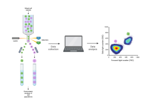
[Flow cytometry schematic. Cells are sent through the flow cytometry instrument one by one. The machine detects their properties and sends them to a computer for analysis. The cells can be separated by type for downstream analysis. Created with BioRender.com.]

Data Analysis
The trainee course I took focused on the step of turning the fluorescent intensity data into something meaningful about different cell populations. The first crucial step is preprocessing, which includes compensation, transformation, and cleaning—basically, the data needs to be tidied up for ease of use. Once this house-keeping task is complete, we can move on to the part we care about, namely identifying the cell populations.

The flow cytometry data can be displayed on a two dimensional plot with each axis representing the fluorescence of a given marker or how much light is scattered in a certain direction. The cells then cluster by type depending on their properties. Traditionally, these cell subpopulations were identified by eye and circled in a process called manual gating. This process can be repeated over and over again comparing different markers giving a complex gating strategy. The gating process can be automated with software packages in R such as FlowDensity and FlowSOM. This strategy looks at density of cells and uses it to determine the cell populations.

[A gating strategy for identifying cell populations. Here we can see the step-wise identification of cell populations, beginning by first identifying live cells, and then distinguishing which cells are lymphocytes from the other cells. From there, the gating strategy separates the cells that are granulocytes and continues finding subpopulations from those that are not granulocytes, and so on, using the properties of the light detected by the flow cytometry machine for each cell. Figures generated in Physalia’s Flow Cytometry Analysis with R/Bioconductor course, 2023.]

Current Research at UH
The Integrative Evolutionary Biology lab, where I am doing my thesis, uses flow cytometry to identify and investigate different types of color cells (or chromophores). We study cichlids which are tropical fish native to lakes and rivers of India, Africa, and South and Central America. There are thousands of different species of these fish with distinct and vibrant color patterns making them an exciting model system to study when it comes to the evolution of cell types and the composition of these fishes’ colorful skin. As shown below, there are a number of different chromatophores: melanophores which are dark in color, iridophores which appear iridescent and blueish, and xanthophores which are yellow. We are using flow cytometry to study the composition of cichlid skin across different species within the Cichlidae family. I’m excited to start applying what I learned in this course to our data.


[Images of the color cells (chromatophores) we are studying in cichlid skin. A) dark dendritic melanophores, B) iridescent, blue iridophores, and C) a close-up of a dendritic yellow xanthophore. Credit Alexandra Faur, Integrative Biology Lab.]

I am grateful to HiLIFE for awarding me the HiLIFE Trainee Conference Grant and to the Integrative Biology Lab for making it possible for me to participate in this excellent course, which deepened my knowledge of flow cytometry, and provided me with the tools to find meaning in this kind of data. I look forward to utilizing this technology in the future.

— Olivia Dreilinger

Reflecting on the journey so far and embarking on a research career

Hello!

This is Rupesh with another blog post to provide an update on what happened during and after my traineeship. You can find my previous post here. It took me a little longer to write this post than promised, but here we are!

Let me start with a short recap: For the traineeship, I worked on my MSc thesis under the supervision of Dr. Ilona Rissanen and Prof. Juha Huiskonen at the Institute of Biotechnology in the University of Helsinki. My thesis was on a project aimed at discovering the structural basis of SARS-CoV-2 neutralization by the antigen-binding fragment of an antibody that was derived from a COVID-19 patient. We can understand how the antibody manages to neutralize the SARS-CoV-2 virus by figuring out where it binds to the spike protein. The antigen-binding fragment, as the name suggests, is the part of an antibody that recognizes and binds to the antigen. In order to find out the epitope of the antigen-binding fragment, I employed single-particle cryogenic electron microscopy (cryoEM). I was able to hypothesize the neutralization mechanism based on the location of the epitope and the subsequent analyses of it.

During the traineeship, I got to learn some cool techniques involved in studying a protein through cryoEM, right from sample preparation to processing the collected data. I would like to highlight a moment during my thesis work, when I got to see how the spike protein looked like in 2D in the early stages of processing the data. It was stunning to see the proteins I had expressed on the computer screen!

But the main takeaways, in my opinion, were the discussions I had with my supervisors and other lab members on how to troubleshoot experiments and interpret the results. That was where I got to understand how to approach science and the problems that arise in these projects. I found these discussions, both formal and informal, to be highly beneficial to my development as a researcher. Writing my thesis was another area where I found myself constantly learning and applying the knowledge to put the big picture together. It involved reading a lot of research articles, which was not an easy task but once I got into the thick of it, I was surprised to notice how fulfilling it was. Staying on top of current research in the field does feel good.

My thesis work had come to an end right after the traineeship period ended and it was time to submit my thesis before the academic year came to a close. As part of graduation requirements, the Genetics and Molecular Biosciences (GMB) master’s program provides the opportunity to present our thesis work during the annual GMB master’s thesis symposium. It was the first time I got to be a part of an in-person symposium in a very long time. In fact, it was the first time I got to see most of my fellow students in the program. All of us presented our work with great enthusiasm and held passionate discussions. It was a good experience to present my results to an audience with varied background and to also listen to talks on different lines of research that my colleagues were working on.

I graduated from the GMB master’s program a couple of months ago and I am incredibly thankful for having the opportunity to work in the research groups of Dr. Rissanen and Prof. Huiskonen. A huge thanks to HiLIFE for supporting me during my thesis. The traineeship resulted in a series of wonderful events which would go on to help me build myself as a researcher and for this, I am grateful!

As for the present, I have recently started to work as a research assistant with the same research groups where I’m hoping to enrich my skills even further before I embark on the journey towards a PhD next year. I’m cherishing this phase of my life where I get to do cool science surrounded by an awesome community, long may it continue!

Cheers,

Rupesh

MANF: no longer a mystery?

Hello everyone! My name is Amanda Sandelin, and I have conducted my traineeship co-supervised by two groups at the University of Helsinki: Mikko Airavaara’s group of BrainRepair and Samuli Ollila’s group of Biophysical Chemistry. If you do not remember what my project was all about, you can refresh your memory here: https://blogs.helsinki.fi/hilife-trainees/2022/07/07/mystery-of-manf/. 

My summer as a HiLIFE research trainee 

My master’s thesis project is about MANF or mesencephalic astrocyte-derived neurotrophic factor. As I wrote in my previous blog post; MANF is a neurotrophic factor, which has protective effects in various disease models, including Parkinson’s disease. MANF mechanisms of action and function are somewhat of a mystery, so this summer I have been working on resolving the mystery of MANF.  

To study the function and structure of MANF, I have been conducting experiments both in vitro and in silico. During my traineeship in Mikko Airavaara’s group I have been working mainly with stem cells. We have a MANF knockout human embryonic stem cell line, which I have been using to study how the loss of MANF affects cellular antioxidant defense. I have also differentiated stem cells into human dopaminergic neurons to study the role of MANF in human dopaminergic neuron development. In Samuli Ollila’s group, we combine molecular dynamics (MD) simulations with nuclear magnetic resonance (NMR) spin relaxation data to study how biomolecules, including proteins, behave. I have been simulating MANF structures in different intracellular conditions using MD simulations and compared the simulations with experimental NMR spin relaxation data to validate the methodology for studying two-domain proteins such as MANF.  

During the summer I have done a lot of cell culturing, MD simulations, immunofluorescent staining, biochemical assays and data analysis. I have had the chance to design, conduct, analyze and discuss experiments and, finally, present my work in seminars and at a conference. So, all around a science-filled summer that has resulted in some exciting results coming your way soon!  

Thank you! 

Did I solve the mystery of MANF? Maybe not yet, but the mission continues, and I have exciting results that I am now compiling in my thesis. The HiLIFE research traineeship made this project possible, and I must thank my supervisors for being open to this collaboration. Thank you to Vassileios Stratoulias, Samuli Ollila and Mikko Airavaara for guiding me as a young researcher and for allowing me to pursue my scientific curiosity.  And, of course, a big thank you to HiLIFE for giving me this opportunity to work in an inspiring research environment!  

Wishing you a cozy autumn,

Amanda Sandelin

Moss mites may tell us about carbon cycling on peatlands

Hi all!

I’m Jemina, a Forest and Atmospheric Sciences student at Helsinki University. This summer I joined Minna Väliranta’s research group at the Environmental Change Research Unit (ECRU) as a HiLIFE Research Trainee. In this blog post, I will briefly introduce you to the hidden life of moss mites. I had the opportunity to investigate a topic new to the whole group and to begin working on my Master’s thesis on the subject.

Moss mite communities may tell us about carbon cycling on peatlands

Organic matter on peatlands is generated and decomposed continuously. Sphagnum moss stems reach up towards the sun while old growth beneath them begins to disintegrate. A mire is formed when growth is faster than decomposition and partially broken-down material compacts into peat. In anoxic conditions under water, peat releases carbon and nutrients very slowly. Peatlands are some of the most efficient carbon banks that nature has to offer. Sometimes, however, decomposition accelerates and exceeds the carbon sequestration of vegetation. This may happen on peatlands modified by human activities or because of changes in climate, but also as a result of the natural development of mires. Whether our peatlands will be carbon sinks or sources in the near future, and how long it will take for possible changes to occur, is a timely topic to research. These different kinds of trajectories are being studied at the University of Helsinki by my supervisor senior research fellow Minna Väliranta and her group.

But how does peat eventually break down? The most effective primary decomposers are microbes, changing the chemical composition of plant matter. Small soil organisms affect decomposition both directly and indirectly: by consuming plant material and other organisms, transporting microbes, and regulating the spread of fungi. Part of the decomposition process depends on the activity of moss mites, a group of tiny insects.


Hard workers underground

Moss mites (Oribatida) are arachnids less than a millimeter in size. Like their better-known relatives, mites have eight legs. Unlike ticks, they are not harmful to humans. On the contrary – without them forestry or food production would be significantly harder. Moss mites play an important role in maintaining the well-being of the earth, as they release nutrients back into the soil. Despite their name, moss mites thrive in a wide variety of environments and are abundant in deserts, anthills, tree foliage, fields, and mires. Hundreds of thousands of moss mites can be found in one square meter of forest soil!

About 12,000 moss mite species are known to the world, and the number is growing constantly as research is carried out. Moss mites can adapt to almost any kind of environment. Some of the species have adjusted to especially harsh conditions, and so mites can also be found in nutrient-poor, acidic and wet peatlands. These environments are demanding for other soil modifiers: for example earthworms cannot be found in peat. This is why moss mites are especially important in peatland environments.

The wide range of species also reflects on the moss mites’ menu. Some species are vegetarians, some enjoy fungal mycelium for food, and others are opportunists eating whatever comes their way. In addition to nutrition, other environmental conditions also affect which moss mite species thrive in each place. Moisture has been found to be an important distinguishing factor, along with soil acidity and phosphorus content.

Knowing moss mite species and their preferences is useful. When any certain species is found, we can deduce that the environmental conditions it requires are also met. Due to their small size, moss mites move very little during their lifetime. It has been noticed that horizontal movement can be just within tens of centimeters. They are also long-lived among insects and can live up to five years in cool habitats. Changes in the species composition in a certain place therefore also reflect a change in the environment. Monitoring of moss mites can show, for example, improving moisture conditions at a restored bog site.

While moss mite communities can tell us about the current state of mires and peatlands, they may also be useful when looking for information of the past.  As peat accumulates, not only plant material but also fossils of small organisms are buried in it. The layered peat deposits of a mire can be dated. By studying the moss mite species at chosen intervals, we can gain information about the environmental conditions that prevailed at the mire tens, hundreds or even thousands of years ago. Moss mites have a hard chitinous shell that survives decomposition quite well. However, small sensory hairs on the mites’ shells may break easily, which often makes it difficult to identify mites to the species level from fossil samples.


Oribatid mites have a hard, chitinous shell and eight legs.


Research is challenging due to the large variety of species

The microscopic size of the organisms and the large variety of species is a challenge in studying moss mites. Determining the species level of mites is hard, and differences between species are sometimes only detectable with the help of DNA research. When I started learning about moss mites, I also noticed that identification guides are poorly available and none of them are in Finnish.

The presence and activity of moss mites has, however, been studied in quite a wide variety of environments in Finland. Riikka Elo noticed in her dissertation that anthills support the life of rich moss mite communities. Inkeri Markkula showed in her work that certain moss mite species in the peat archives can tell us if permafrost has previously been present at northern mires. Ritva Penttinen, now retired, has had a long career in tick and mite research at the Turku University Zoological Museum, and has extensively mapped Finnish moss mite species.

In planning my own research, I have been most interested in the question of whether the moss mite community could tell us something about the rate of soil carbon decomposition. Since mites stay put in a very small area, the species composition can significantly change within the scale of just a few meters. By collecting moss samples from different kinds of mire surfaces and separating the mites from them, my intention is to compare the found species and their abundances with carbon flux measurements from each location. Doing research is always an adventure, and I still can’t be sure what I’ll find along the way! I will keep you posted as the research continues.