Reflecting on the journey so far and embarking on a research career

Hello!

This is Rupesh with another blog post to provide an update on what happened during and after my traineeship. You can find my previous post here. It took me a little longer to write this post than promised, but here we are!

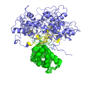
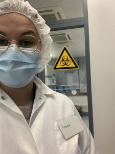
Let me start with a short recap: For the traineeship, I worked on my MSc thesis under the supervision of Dr. Ilona Rissanen and Prof. Juha Huiskonen at the Institute of Biotechnology in the University of Helsinki. My thesis was on a project aimed at discovering the structural basis of SARS-CoV-2 neutralization by the antigen-binding fragment of an antibody that was derived from a COVID-19 patient. We can understand how the antibody manages to neutralize the SARS-CoV-2 virus by figuring out where it binds to the spike protein. The antigen-binding fragment, as the name suggests, is the part of an antibody that recognizes and binds to the antigen. In order to find out the epitope of the antigen-binding fragment, I employed single-particle cryogenic electron microscopy (cryoEM). I was able to hypothesize the neutralization mechanism based on the location of the epitope and the subsequent analyses of it.

During the traineeship, I got to learn some cool techniques involved in studying a protein through cryoEM, right from sample preparation to processing the collected data. I would like to highlight a moment during my thesis work, when I got to see how the spike protein looked like in 2D in the early stages of processing the data. It was stunning to see the proteins I had expressed on the computer screen!

But the main takeaways, in my opinion, were the discussions I had with my supervisors and other lab members on how to troubleshoot experiments and interpret the results. That was where I got to understand how to approach science and the problems that arise in these projects. I found these discussions, both formal and informal, to be highly beneficial to my development as a researcher. Writing my thesis was another area where I found myself constantly learning and applying the knowledge to put the big picture together. It involved reading a lot of research articles, which was not an easy task but once I got into the thick of it, I was surprised to notice how fulfilling it was. Staying on top of current research in the field does feel good.

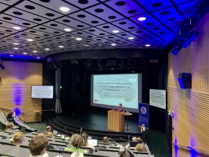
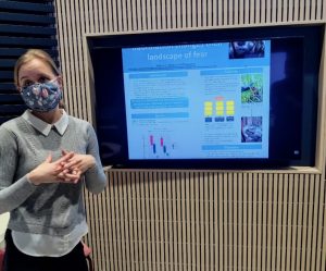
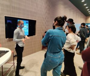
My thesis work had come to an end right after the traineeship period ended and it was time to submit my thesis before the academic year came to a close. As part of graduation requirements, the Genetics and Molecular Biosciences (GMB) master’s program provides the opportunity to present our thesis work during the annual GMB master’s thesis symposium. It was the first time I got to be a part of an in-person symposium in a very long time. In fact, it was the first time I got to see most of my fellow students in the program. All of us presented our work with great enthusiasm and held passionate discussions. It was a good experience to present my results to an audience with varied background and to also listen to talks on different lines of research that my colleagues were working on.

I graduated from the GMB master’s program a couple of months ago and I am incredibly thankful for having the opportunity to work in the research groups of Dr. Rissanen and Prof. Huiskonen. A huge thanks to HiLIFE for supporting me during my thesis. The traineeship resulted in a series of wonderful events which would go on to help me build myself as a researcher and for this, I am grateful!

As for the present, I have recently started to work as a research assistant with the same research groups where I’m hoping to enrich my skills even further before I embark on the journey towards a PhD next year. I’m cherishing this phase of my life where I get to do cool science surrounded by an awesome community, long may it continue!

Cheers,

Rupesh

MANF: no longer a mystery?

Hello everyone! My name is Amanda Sandelin, and I have conducted my traineeship co-supervised by two groups at the University of Helsinki: Mikko Airavaara’s group of BrainRepair and Samuli Ollila’s group of Biophysical Chemistry. If you do not remember what my project was all about, you can refresh your memory here: https://blogs.helsinki.fi/hilife-trainees/2022/07/07/mystery-of-manf/. 

My summer as a HiLIFE research trainee 

My master’s thesis project is about MANF or mesencephalic astrocyte-derived neurotrophic factor. As I wrote in my previous blog post; MANF is a neurotrophic factor, which has protective effects in various disease models, including Parkinson’s disease. MANF mechanisms of action and function are somewhat of a mystery, so this summer I have been working on resolving the mystery of MANF.  

To study the function and structure of MANF, I have been conducting experiments both in vitro and in silico. During my traineeship in Mikko Airavaara’s group I have been working mainly with stem cells. We have a MANF knockout human embryonic stem cell line, which I have been using to study how the loss of MANF affects cellular antioxidant defense. I have also differentiated stem cells into human dopaminergic neurons to study the role of MANF in human dopaminergic neuron development. In Samuli Ollila’s group, we combine molecular dynamics (MD) simulations with nuclear magnetic resonance (NMR) spin relaxation data to study how biomolecules, including proteins, behave. I have been simulating MANF structures in different intracellular conditions using MD simulations and compared the simulations with experimental NMR spin relaxation data to validate the methodology for studying two-domain proteins such as MANF.  

During the summer I have done a lot of cell culturing, MD simulations, immunofluorescent staining, biochemical assays and data analysis. I have had the chance to design, conduct, analyze and discuss experiments and, finally, present my work in seminars and at a conference. So, all around a science-filled summer that has resulted in some exciting results coming your way soon!  

Thank you! 

Did I solve the mystery of MANF? Maybe not yet, but the mission continues, and I have exciting results that I am now compiling in my thesis. The HiLIFE research traineeship made this project possible, and I must thank my supervisors for being open to this collaboration. Thank you to Vassileios Stratoulias, Samuli Ollila and Mikko Airavaara for guiding me as a young researcher and for allowing me to pursue my scientific curiosity.  And, of course, a big thank you to HiLIFE for giving me this opportunity to work in an inspiring research environment!  

Wishing you a cozy autumn,

Amanda Sandelin

Moss mites may tell us about carbon cycling on peatlands

Hi all!

I’m Jemina, a Forest and Atmospheric Sciences student at Helsinki University. This summer I joined Minna Väliranta’s research group at the Environmental Change Research Unit (ECRU) as a HiLIFE Research Trainee. In this blog post, I will briefly introduce you to the hidden life of moss mites. I had the opportunity to investigate a topic new to the whole group and to begin working on my Master’s thesis on the subject.

Moss mite communities may tell us about carbon cycling on peatlands

Organic matter on peatlands is generated and decomposed continuously. Sphagnum moss stems reach up towards the sun while old growth beneath them begins to disintegrate. A mire is formed when growth is faster than decomposition and partially broken-down material compacts into peat. In anoxic conditions under water, peat releases carbon and nutrients very slowly. Peatlands are some of the most efficient carbon banks that nature has to offer. Sometimes, however, decomposition accelerates and exceeds the carbon sequestration of vegetation. This may happen on peatlands modified by human activities or because of changes in climate, but also as a result of the natural development of mires. Whether our peatlands will be carbon sinks or sources in the near future, and how long it will take for possible changes to occur, is a timely topic to research. These different kinds of trajectories are being studied at the University of Helsinki by my supervisor senior research fellow Minna Väliranta and her group.

But how does peat eventually break down? The most effective primary decomposers are microbes, changing the chemical composition of plant matter. Small soil organisms affect decomposition both directly and indirectly: by consuming plant material and other organisms, transporting microbes, and regulating the spread of fungi. Part of the decomposition process depends on the activity of moss mites, a group of tiny insects.


Hard workers underground

Moss mites (Oribatida) are arachnids less than a millimeter in size. Like their better-known relatives, mites have eight legs. Unlike ticks, they are not harmful to humans. On the contrary – without them forestry or food production would be significantly harder. Moss mites play an important role in maintaining the well-being of the earth, as they release nutrients back into the soil. Despite their name, moss mites thrive in a wide variety of environments and are abundant in deserts, anthills, tree foliage, fields, and mires. Hundreds of thousands of moss mites can be found in one square meter of forest soil!

About 12,000 moss mite species are known to the world, and the number is growing constantly as research is carried out. Moss mites can adapt to almost any kind of environment. Some of the species have adjusted to especially harsh conditions, and so mites can also be found in nutrient-poor, acidic and wet peatlands. These environments are demanding for other soil modifiers: for example earthworms cannot be found in peat. This is why moss mites are especially important in peatland environments.

The wide range of species also reflects on the moss mites’ menu. Some species are vegetarians, some enjoy fungal mycelium for food, and others are opportunists eating whatever comes their way. In addition to nutrition, other environmental conditions also affect which moss mite species thrive in each place. Moisture has been found to be an important distinguishing factor, along with soil acidity and phosphorus content.

Knowing moss mite species and their preferences is useful. When any certain species is found, we can deduce that the environmental conditions it requires are also met. Due to their small size, moss mites move very little during their lifetime. It has been noticed that horizontal movement can be just within tens of centimeters. They are also long-lived among insects and can live up to five years in cool habitats. Changes in the species composition in a certain place therefore also reflect a change in the environment. Monitoring of moss mites can show, for example, improving moisture conditions at a restored bog site.

While moss mite communities can tell us about the current state of mires and peatlands, they may also be useful when looking for information of the past.  As peat accumulates, not only plant material but also fossils of small organisms are buried in it. The layered peat deposits of a mire can be dated. By studying the moss mite species at chosen intervals, we can gain information about the environmental conditions that prevailed at the mire tens, hundreds or even thousands of years ago. Moss mites have a hard chitinous shell that survives decomposition quite well. However, small sensory hairs on the mites’ shells may break easily, which often makes it difficult to identify mites to the species level from fossil samples.


Oribatid mites have a hard, chitinous shell and eight legs.


Research is challenging due to the large variety of species

The microscopic size of the organisms and the large variety of species is a challenge in studying moss mites. Determining the species level of mites is hard, and differences between species are sometimes only detectable with the help of DNA research. When I started learning about moss mites, I also noticed that identification guides are poorly available and none of them are in Finnish.

The presence and activity of moss mites has, however, been studied in quite a wide variety of environments in Finland. Riikka Elo noticed in her dissertation that anthills support the life of rich moss mite communities. Inkeri Markkula showed in her work that certain moss mite species in the peat archives can tell us if permafrost has previously been present at northern mires. Ritva Penttinen, now retired, has had a long career in tick and mite research at the Turku University Zoological Museum, and has extensively mapped Finnish moss mite species.

In planning my own research, I have been most interested in the question of whether the moss mite community could tell us something about the rate of soil carbon decomposition. Since mites stay put in a very small area, the species composition can significantly change within the scale of just a few meters. By collecting moss samples from different kinds of mire surfaces and separating the mites from them, my intention is to compare the found species and their abundances with carbon flux measurements from each location. Doing research is always an adventure, and I still can’t be sure what I’ll find along the way! I will keep you posted as the research continues.

Crossing Continents: The journey of a budding neuroscientist

Hi everyone! My name is Anushka Wakade. In this blog, I am going to share
my journey from being just a budding neuroscience enthusiast to an
international HiLIFE trainee in the Neuroscience Center of Helsinki.

A Peek into my Life before Finland

As far back as I can remember, the workings of the human brain have
always fascinated me. The intricacies of human behavior and the fallout
that is seen when the brain glitches has never failed to intrigue me. As a
result, even as a bachelor’s student in life-sciences, I have tried my best to
go out of my way and read and obtain as much experience as I could on
this topic. During my extra honor courses and internships, I realized that
neurodegeneration, cognition and the link between the two appealed to me in this massively broad field of neurosciences. Keeping in mind that the next
logical step was to pursue my further education (as a master’s degree) in
the same, I started looking for master’s courses that were offering similar
experience in Europe (as neuroscience related research is expanding here
exponentially) . The master’s program in the University of Helsinki fit all my
expectations with respect to courses, practical experiences and exposure
to opportunities. Hence, it was not a difficult decision to finalize Helsinki
and move here for my further education.

Perks of being a HiLIFE Trainee

During my time as a HiLIFE trainee. I decided to join as a trainee in Dr.
Coralie Di Scala’s group in the Neuroscience Center. The lab is focused on
studying the lipid-protein interactions in nervous system diseases, with a
special emphasis on epilepsy. Having studied biochemistry extensively
during my bachelor’s, my interests and suitability to work in a group which
studies neurodegeneration from a biochemical point of view worked out
well for me to decide that I wanted to pursue my master’s thesis in the
same lab.
Temporal Lobe Epilepsy is one of the most common types of epilepsy
whose clinical definition is the presence of unprovoked recurrent seizures
over a period longer than 24 hours. Approximately 30 to 50% of epileptic
patients suffer from severe cognitive deficits (such as memory alterations)
whose severity only increases the longer they have seizures. Being one of
the most globally prevalent disorders, research concerning its
pathophysiology has been under work for a couple of decades now.
Unfortunately, no cure or aetiology has been identified so far. The current
anti-epileptic drugs used to counteract the intense symptoms have been
found to be ineffective in about one third of the epileptic patients. As a
result, the condition of epileptic patients continues to worsen, decreasing
their quality of life. For this reason, it is crucial to direct research towards
discovering the underlying mechanism of epilepsy, which would lead to
finding more effective therapies. Currently, there are quite a few theories
investigating this underlying molecular mechanism and one of the most
widely accepted for Temporal Lobe epilepsy is that the alteration of
chloride ion homeostasis in neurons causes a grave impact on the
synaptic (neuronal) communications in the brain, precipitating the
apparition of seizures. The disturbances in neuronal chloride homeostasis
in epilepsy are caused by the malfunctioning of the chloride ion
co-transporters present on the cell membrane, which are responsible for regulating the ionic flux between cells. Our laboratory is interested in exploring the missing or the unknown aspects of this specific theory. Dr. Di Scala’s lab has discovered that certain lipids present in the cellular membrane (Gangliosides) have specific and direct
interactions with these chloride cotransporters and hence also plays a
crucial role in maintaining it’s structural and functional integrity,
necessary for the normal neuronal functioning. Despite knowing the other
vital functions of gangliosides, no other research group has explored it’s role in
the pathophysiology of epilepsy and its potential therapies. In this way, the
laboratory is novel in its approach and explores avenues that have not yet
been taken into account for explaining the mechanism and progression of
this disease through modulation of these chloride transporters. This would open several avenues
for new therapies concerning epilepsy. The lab is currently focused on quantifying and then studying the interactions between these gangliosides (membrane lipid) and protein (chloride transporter) to get a fuller picture of the pathophysiology of epilepsy.

My thesis would be a sub-project of this larger research question
that the laboratory is tackling by focusing on the characterization of lipid
alteration during epilepsy.

I have been working on this for a couple of months but I still have a long
way to go before I wrap up my thesis. I am extremely excited and hopeful
for the results that will start rolling in soon. During this period, I have not
just grown as a researcher but also as a person. The support and
teachings by both my supervisors have been invaluable and I am sure they
will stay with me wherever I go next in my career! I will elaborate more on
this and my future plans in my next post once I finish my  thesis. Thank you for being interested! I will keep you updated!

Behind the Mind


Hi everyone!

I’m Santeri Lepistö, a HiLIFE research trainee and master’s student in the neuroscience programme at the University of Helsinki. I carry out my HiLIFE internship in Satu Palva’s research group, focusing on studying the link between human brain dynamics and psychological processes. In this blog post, I give an introduction to my academic interests and internship’s research topic.


Brain-Mind Relationship

Before studying neuroscience I graduated in psychology from the University of Jyväskylä and worked for a while as a psychologist conducting neuropsychological assessments for children with neurological disorders and developmental delay. What especially fascinates me in science is the brain-mind relationship: how the brain – in a close interaction with the rest of the body – computes mental phenomena. At the level of nervous system, I’m intrigued by the human brain as a complex information-processing device that widely regulates physiological and behavioral functions. At the level of mind and behavior, my interests incline to basic psychological processes like cognitive functions, emotions, learning and development. It makes me wonder, for instance, how attention can be considered as “the set of evolved brain processes that leads to adaptive and effective behavioral selection” [1] and how, on the other hand, emotions have been proposed to serve as a coordinating mechanism, mode of operation that adjusts states of the brain and body influencing thoroughly on individual’s way to perceive, think and behave [2].


Brain, Mind and the Big Picture

My urge to examine brain-mind relationship is inspired by the puzzling questions regarding origins of the brain, mind and behavior. In order to understand astonishingly complicated human condition in all its neuronal capacity and constraints, it is important, in my opinion, to combine knowledge from both evolutionary and cultural foundations as an integrative evolutionary-cultural framework. In other words, to put multifaceted emphasis on where we come from and where we’re living. We are, as a species, an outcome of monumental evolutionary history and possess, among other organs, a brain that is shaped by evolutionary processes, such as natural selection. Alongside of acknowledging our evolutionary past, genetic makeup and biologically grounded predispositions, it is essential to underline the impact of cultural context on human ontogeny and daily living. In addition, it is pivotal to pay attention to altered environmental demands that occur in the modern world in contrast to ancestral ecological niche. Today, our brains and minds interact with factors like technology, city life, advanced medicine, educational system, art and literature, governmental policy, science and HiLIFE blog posts. The high degree coordination of human cultural practices can be traced back to brain and cognition, namely, our species-specific neurocognitive capability to establish shared goals and accumulate knowledge over time [3]. Moreover, we have an impressive ability to learn – brain plasticity to form internal models of external world [4]: humans not only use object recognition to detect faces, spoken language to communicate ideas and social cognition to cooperate but also harness their brain circuits to acquire sophisticated cultural skills concerning man-made inventions like, in case of writing systems, learn to read [5] by recognizing written words, decoding meaning of a text and taking someone else’s perspective in a novel. 


Inside the Brain: Oscillatory Activity 

Indeed, humans have an exceptional track record of peculiar cultural practices. But how the brain, more specifically, manages to orchestrate these kinds of complex patterns of behavior? During the HiLIFE trainee period I explore how the brain computes the mind through the lens of systems and cognitive neuroscience – by investigating activity of large-scale neuronal networks and its association to different psychological processes. When observing the beauty of the natural world, rhythms and synchrony can be found in several places, of which one is the human brain. Collective action of neurons generate rhythmic electrophysiological activity that can be studied by using brain imaging techniques like magnetoencephalography, a tool I use during my research trainee period. These oscillations, electrical ups and downs produced by vast neuronal populations, are considered to vividly reflect how the brain selects, modifies and transmits information. 

To elaborate, brain’s information transmission from one place to another can be addressed by the concept called functional or effective connectivity – describing the correlation or dependence of neuronal activity from each other. Functional and effective connectivity are influenced by the structure of the brain and connect areas with similar functions. According to the hypothesis called communication-through-coherence, selective information transmission occurs when oscillations in two brain regions are synchronized and act coherently [6]. This synchronization, which is a central research topic in systems and cognitive neuroscience, provides an enlightening window to understand brain-mind relationship. For example, brain synchronization has been previously linked to attentional capacity, that is how many objects one can attend concurrently. The study conducted by Palva and colleagues suggests that individual attentional capacity is dependent on how the brain succeeds to integrate activity of different high frequency oscillations in large-scale neuronal networks [7]. From this perspective, I think the research of system-level human brain dynamics serves as an invigorating way to find novel questions and answers in the search for what’s the story behind the mind. 

Magnetoencephalography raw data showing oscillations from the human brain (in a time period of five seconds)


In the next blog post, I will tell you more about my
HiLIFE research trainee experience!


References 

  1. Krauzlis, R. J., Wang, L., Yu, G., & Katz, L. N. (2021). What is attention?. Wiley Interdisciplinary Reviews: Cognitive Science, e1570.
  2. Al-Shawaf, L. (2021, December 28). What Are Emotions?. Psychology Today. https://www.psychologytoday.com/us/blog/six-impossible-things-breakfast/202112/what-are-emotions.
  3. Tomasello, M. (2019). Becoming human. In Becoming Human. Harvard University Press.
  4. Dehaene, S. (2020). How we learn: The new science of education and the brain. Penguin UK. 
  5. Dehaene, S. (2009). Reading in the brain. New York. 
  6. Fries, P. (2015). Rhythms for cognition: communication through coherence. Neuron, 88(1), 220-235. 
  7. Rouhinen, S., Siebenhühner, F., Palva, J. M., & Palva, S. (2020). Spectral and anatomical patterns of large-scale synchronization predict human attentional capacity. Cerebral Cortex, 30(10), 5293-5308. 

Six Months after Country Hopping to Switzerland: Master’s thesis is submitted!

Hello again everybody!

This is Rosa López again updating you about my internship as a HiLIFE Research Trainee! As you may remember (or not, please click here for the previous blog post 🙂 ) I used this scholarship to carry out my Master’s thesis as an international student at EPFL located in Lausanne (Switzerland). After six months of tough learning and hard work, I am delighted to inform you that my thesis is finished! In this blog post, I want to explain to you how the whole process ended, from the first experiments to the thesis submission. So, let’s dig in!

What was my thesis about? How was it performed?

As a quick recap of the previous blog post ( 😀 ), my Master’s thesis was performed at the Laboratory of Stem Cell Bioengineering (LSCB)1. This lab, led by Prof. Matthias Lütolf, aims to develop third-generation organoids from stem cells by using innovative bioengineering strategies. One of the research lines focuses on the development of homeostatic human gastric third-generation organoids from human biopsies since this current organoid model contains several limitations2. This project is being conducted by Moritz Hofer, a PhD student in LSCB and my supervisor throughout my whole thesis. So, you may ask: Rosa, what was your Master’s thesis about?

The main aim of my thesis was to test the effect of different extracellular matrix (ECM) proteins on gastric stem cell differentiation. The ECM is currently considered one of the key stem cell niche components3,4. As a matter of fact, several ECM proteins had been already established to have a specific location within the human gastric mucosa. Thus, we wanted to check if these proteins could influence stem cell maintenance or differentiation towards one specific cell type. The main workflow of the project was to seed gastric organoid-derived epithelial cells on the proteins of interest and check for stem cell markers or other gastric epithelial cell markers with quantitative polymerase chain reaction (qPCR) after some time.

However, an important question arose right at the beginning of the project: how to perform this experiment? Some solutions could have been coating the seeding plate with the ECM protein of interest or using a mixture of Matrigel® with our ECM protein of interest. However, the former solution did not resemble the biomechanics of the mucosa, whereas the Matrigel® meant a too complex and uncontrollable environment. That is the reason why we decided to use synthetic hydrogels5, whose biomechanical properties can be modelled, and they are enriched only with our ECM proteins of interest. Even though it was a straightforward solution, a major part of the thesis was the bioengineering of synthetic hydrogel. The whole optimization took more than half of the internship! In the end, we were able to obtain preliminary results which showed that indeed some ECM proteins maintain stem cells, whereas others enhance differentiation towards other gastric cell types.

After all the experiments were done there was still one part missing… The whole thesis writing. I guess I am like most students, leaving all the writing towards the end. I would advise you to not do it. Although probably you’ll do the same mistake, so if you’re at that stage at this very moment… good luck!

And now, what is the next chapter?

My internship went on for six months and after I submitted my thesis, I got the Master’s graduation. During this time, I reassure myself what I want to do next: PhD. I still do not know where, but I know for sure that I like stem cell research. In the next months, I will be doing another internship before the PhD focused on the scarring and repair in the central nervous system at Karolinska Institutet. Let’s see how that goes and how it affects my future!


I would like to finish this post by acknowledging all the LSCB team, and specifically thanking Moritz Hofer for all his help and mentoring during this internship. I believe that having good mentoring is essential for success! Also, to all the people that were with me during the whole process, either physically in Switzerland or through the phone. Emotional support is more than necessary to complete a good thesis. Last but not least, thanks to Switzerland for having such breathtaking nature and landscapes. Although long hours in the lab are necessary, having some getaways is as important. I will bless you with some Swiss pics down below 😀

Thank you all for reading! Hope the best for you :3

Bests,

Rosa


Some (maybe) interesting links: 

  1. Laboratory of Stem Cell Bioengineering Webpage: https://www.epfl.ch/labs/lutolf-lab/
  2. Seidlitz, T., Koo, B. K., & Stange, D. E. (2020). Gastric organoids—an in vitro model system for the study of gastric development and road to personalized medicine. Cell Death & Differentiation, 28(1), 68–83. https://doi.org/10.1038/s41418-020-00662-2
  3. Pardo-Saganta, A., Calvo, I. A., Saez, B., & Prosper, F. (2019). Role of the Extracellular Matrix in Stem Cell Maintenance. Current Stem Cell Reports 2019 5:1, 5(1), 1–10. https://doi.org/10.1007/S40778-019-0149-9
  4. Rezakhani, S., Gjorevski, N., & Lutolf, M. P. (2021). Extracellular matrix requirements for gastrointestinal organoid cultures. Biomaterials, 276. https://doi.org/10.1016/J.BIOMATERIALS.2021.121020
  5. Madduma-Bandarage, U. S. K., & Madihally, S. V. (2021). Synthetic hydrogels: Synthesis, novel trends, and applications. Journal of Applied Polymer Science, 138(19), 50376.  https://doi.org/10.1002/APP.50376

Mystery of MANF

Hello everyone! My name is Amanda Sandelin, and I am a first-year (soon to be second-year) Master’s student in Translational Medicine. I am one of the HiLIFE Research Trainees of 2022, and I am conducting my traineeship co-supervised by two groups at the University of Helsinki; Mikko Airavaara’s group of Neuroprotection and Neurorepair and Samuli Ollila’s group of Biophysical Chemistry. My interest lies in neuroscience, but I am also interested in structural biology as a tool to help understand the details of what really is happening in our brains.  

The star of my project: MANF 

My projects revolve heavily around one protein, namely the mesencephalic astrocyte-derived neurotrophic factor or, easier said, MANF. Even though MANF is a neurotrophic factor, its characteristics differ significantly from other “traditional” neurotrophic factors. In fact, the mechanisms of action and functions of MANF are still quite a mystery. But why are we interested in this one protein? Well, what is known about MANF is that it has pleiotropic protective effects in various disease models, including Parkinson’s disease, and it is important in human development. By studying the mechanisms of MANF, we can better understand neuroprotection and identify possible new therapeutic targets. 

The next question is of course: how do we study this?  I work both in vitro and in silico, which means I work with cells and by computational models, more specifically human embryonic stem cells and molecular dynamic (MD) simulations.  Vassileios Stratoulias in our lab has established a protocol for differentiation of both wildtype and MANF knockout stem cells into dopaminergic neurons based on a previously published rigorous protocol. Using this setup, we can study the differences between wildtype and MANF knockout cells at different stages of development. In Samuli Ollila’s group, we use MD simulations and NMR to look at MANF on the molecular level and see if different conditions (such as different pH, ATP or ion concentrations) affect the structure and function of MANF.  

My first month 

I have been loving the first month of my traineeship, and I have already had a chance to learn a lot of things and immerse myself in science and research. I have been doing a lot of cell culturing, simulations, experiments, planning, analysis, and discussing and I even attended a conference, where I got to present a poster. Below are some images to really convey the amazing, science-filled month I have been enjoying. Thank you goes out to my supervisors, Vassileios Stratoulias, Samuli Ollila and Mikko Airavaara, and to everyone else in the groups for making my traineeship as great as it is. And, of course, a big thank you to HiLIFE for giving me this opportunity to experience research at its heart! 

Here you can read more about: 

Neuroprotection and neurorepair 

Biophysical chemistry 

Wishing you a lovely summer,  

AmandaSandelin


Meet the Aalto-Helsinki iGEM team 2022!

Hello! We are the Aalto-Helsinki iGEM team 2022 and here is a little bit about us and about our project.

First things first, what is iGEM?

iGEM stands for “international genetically engineered machine” and is a global synthetic biology competition between more than 350 teams around the world. We are a team of 10 highly-motivated students from both Aalto University and the University of Helsinki. The combined Aalto-Helsinki team has existed since 2014, but the team members are different every year. Last February, the 2021 Team chose us as the new members for the coming year, and since then we have been brainstorming about what our project should be. 

Every year, the team picks a research idea and spends the summer break implementing it in the lab, and then presenting the results at the Grand Jamboree (which this year will take place in Paris in October). As you can see, we have a very short time frame to organize, visualize, and prove our idea!


What is our idea?

After a lot of back-and-forth between several promising ideas, we decided to target biofilms on chronic wounds. Chronic wounds are found in 15 % to 25 % of diabetes patients and often lead to increased morbidity, mortality in general decreasing the quality of life, and are therefore of high public health concern. 

The environment of chronic wounds is usually low in oxygen (hypoxic) and thereby causes decreased immune activity. Therefore, bacteria can easily settle there, leading to the formation of biofilms. Biofilms are structural communities of bacteria that are usually tolerant to host defences and antibiotics. This is an issue for the patient, because treatment is more difficult.

Biofilms form when bacteria settle on the wound site, and their presence attracts even more bacteria through a process called quorum sensing. Quorum sensing is defined as population density measurement and a form of inter-microbial communication in the biofilm. The process of quorum sensing specifically works through bacteria releasing small molecules or peptides that other bacteria can take up, thereby sensing the presence of the other bacteria. Furthermore, bacteria can also auto-induce themselves via these peptides or molecules: indeed, auto-induction is a positive feedback-loop in which bacteria signal to themselves to produce even more of these quorum sensing peptides or molecules, through which further downstream gene activity and more biofilm build-up is achieved.

Our central aim is to target this mechanism to disrupt the formation of more biofilms.

For this we want to utilize DARPins. DARPin stands for designed ankyrin repeat proteins. They are genetically engineered peptides that mimic antibodies. We want to design DARPins that bind to the quorum sensing peptides released by the bacteria, which should prevent the “communication” between bacteria and obstruct the further build-up of biofilm. The end result we envision is for our designed DARPins to be used in combinatorial therapy with antimicrobial agents against biofilms, as blocking their signalling should make the bacteria more sensitive to such antimicrobials. 


Is that all?

In fact, iGEM is a lot more than just the research aspect. In addition to our main project, we also focus on developing different collaborations, as well as community outreach and science communication. The latter aspect is called Human Practices and represents a key foundation of the iGEM competition. As part of this, we are currently talking to many different experts in the field, including front-line medical staff, and are very happy to learn more about the practical considerations of wound healing. We will also prepare and hold a workshop at Heureka, and collaborate with The Science Basement. We plan to give a talk on the 24th of September during the European Biotech Week. We furthermore continue the Aalto-Helsinki iGEM podcast that was established last year.

So, if you want to know how everything is going throughout our project, you can follow us there or visit our blog.


Thank you for reading and thank you to HiLIFE!

Lastly, we also want to thank HiLIFE and the University of Helsinki for supporting us and our idea during the iGEM program and for enabling us to join this competition. We are very happy to be part of iGEM and cannot wait to get started, working to make our theories a reality! 

When puberty hits you

Hi all,

My name is Linda Helena Müller and I am writing to update you on my HiLIFE Research Traineeship. During the last six months, I have been working on puberty research in the Raivio group at Helsinki University. The time flew by and I am excited to have successfully finished my Master’s thesis project recently. In fact, I just submitted my thesis last month. I used the HiLIFE scholarship to explore a field of research I have not been in contact with before. In my project, I worked in the area of stem cell research and neuroscience. Specifically, I have used the CRISPR/Cas9 system to activate a gene associated with puberty initiation. The traineeship allowed me to improve my skills in the fields of genetics and cell culture. However, I have also learned a lot about other stem cell research areas by attending talks and a retreat of the Stem Cells and Metabolism Research Program at Helsinki University. I am glad to report that the HiLIFE Traineeship completely fulfilled its purpose of exploring a research curiosity of mine.

Being at the end of my Master’s degree, I am now sure that I want to keep following the academic career and enroll in a PhD program after graduation. This traineeship and the methods I have learned were a tremendous help in getting into the PhD program of my choice. I will start my PhD at the EMBL institute later this year and continue in the same research field. Therefore, I am highly thankful to have been chosen as one of the HiLIFE trainees. This research period greatly helped me to orientate on what field I would like to keep working in and allowed me to learn important methods for doing so.

Aside from the academic part, performing this traineeship also gave me the opportunity to further experience life in Helsinki. I have fallen in love with the city and was thrilled to attend this year’s Vappu celebration since it was canceled last year. I even fried Munkit with the help of a Finnish friend. I also added my favorite picture of the Helsinki city center which I took on the night of the Lux light festival – check it out below. I am sad to leave Finland, however, the last months have been an excellent ending to my Master’s studies in Helsinki.

Presenting your own work allows a chance to network!

What an experience!

Truly the HiLIFE traineeship period has given me so much, not only in experiences but in chances to grow and develop as a person. I have managed to collect behavioural data on reed warbler incubation (see my previous post, on to fear or not to fear) and submit my thesis for evaluation. Although in the researching world there is always something that can be polished off and rewritten, I am confident in the quality and standard of my work, thanks to the invaluable help from my supervisor and the research team. What amazed me the most was how helpful and willing others were to answer my questions, take time off their busy schedule to help me and provide me with constructive critique that helped me develop as a young researcher. These last months have left me a lot richer in skills and experiences.

Me presenting my poster at the Spring Symposium 2022.

During the last months, I had the chance to present my work in several varying setting with a changing audience and style of presentation. Although I managed to create interactive and engaging power points targeting different audiences, I must say the highlight was the poster session I got to attend. This was a part of the LUOVA Spring Symposium, organized by doctoral students, focusing on the research in ecology, evolutionary biology, and conservation. Here I presented my work to a wide audience, from fellow students to foreign researchers. The questions and constructive feedback I received helped me dive deeper into my work, and I could see new ideas taking shape through conversations I had with others. The cherry on top of the cake was that this gave me the opportunity to form new connections and network in the researching world, as well as glimpse at the current biology research that is carried out at the University of Helsinki.

Discussing scientific work with others gives a chance to hear other ideas on the same topic.

Although research is very individual based work and stems from personal interests, I learned the importance of sharing differing perspectives and ideas through conversation. As one starts to dive deeper into a topic, it may be harder to see the broader questions that arise from the work at hand. Discussing with others and brainstorming the overall impact of the topic provides a broad umbrella that allows the work to be applicable for several different research questions, as well as allows others to take the key aspects into account within their own research. This forms a web of support for the current research at hand. I am thankful to have been a part of a research group with individuals from various backgrounds, that provided diverse ideas and opinions that helped me build my research into the landscape of fear concept. As a young researcher, the help and support of experienced researchers is critical to navigate the field full of emerging questions. An extra special thank you for this to my supervisor, Rose Thorogood.

A couple of days ago an old professor told me that a few decades ago, research, particularly in birds, used to be dominated by one researcher. This meant that one researcher specialized on one species, and it was frowned upon if somebody wanted to study that species individually. I was surprised at this, and we had a very interesting discussion on why this was the case. We both agreed that research becomes richer the more people look at a similar species (or question) from their own personal angle. This provides more ideas and opportunities to form a diverse understanding of why we see what we see in nature. I really feel that my traineeship has allowed me to see this richness through working in a research group.

Having had the chance to watch experts within their fields navigate research topics, I realize there is a vast ground of knowledge to be consumed. I feel that some of these skills can be best gained in the working world, to understand what data is already existing and waiting to be analysed and pondered upon. We are very lucky to live in a society where the government supports museums and the upkeep of long-term data sets. However, these need to be actively utilized and inspected to determine what type of research is most beneficial for conserving nature and the ecosystem. I am ever so thankful for the HiLIFE traineeship that has supported me and my journey in experiencing the researching world. I hope that in my future, I can continue in the researching world and maybe even provide support to other young researcher someday, as I have been supported by the professors at the University of Helsinki and financially by HiLIFE.