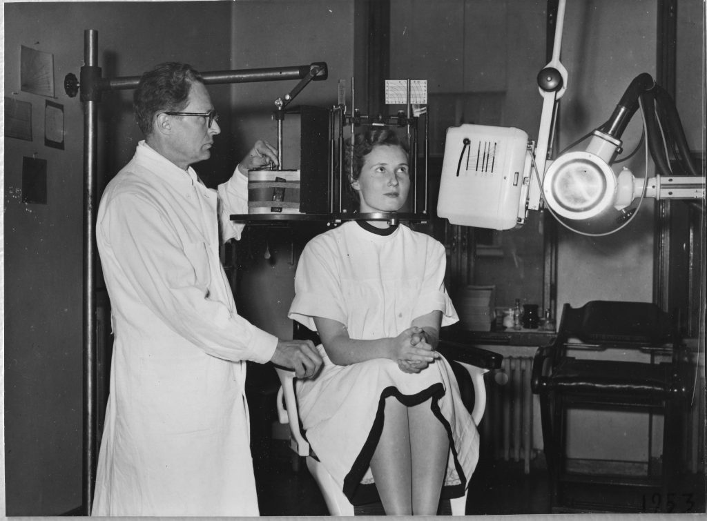
After the end of the Second World War in 1945, Yrjö V. Paatero, Doctor of Dental Science, worked at the University of Helsinki’s Institute of Dentistry, overseeing X-ray examinations and diagnostics. He longed to do research work, but had little time for it because his days were filled with routine tasks. At the time, dental X-rays were taken by placing a film in the patient’s mouth time and again because several X-rays had to be taken to determine the condition of the entire dentition. Paatero was keen to find a less time-consuming solution, and the seed of the idea of panoramic radiography began to germinate. However, the road to this point had been far from simple, and several stumbling blocks still remained.
Long road to an academic research career
Yrjö Veli Paatero was born in 1901 in Helsinki. In 1910, his family moved to a farm in the North Savo region of eastern Finland. He was due to continue his schooling in Kuopio, but the difficulties that emerged as a result of the change of school and the homesickness he felt when living in a lodging house put a temporary end to the then 10-year-old Yrjö’s school days. He did not continue his studies until 1916 when the family returned to Helsinki. Yrjö first studied metalwork at a preparatory vocational school for boys and then completed his lower secondary education while working as a stockkeeper. He took the matriculation examination in 1933 at the age of 32, following studies at the evening school of the Finnish Private Lyceum.
He immediately embarked on studies in dentistry, all the while continuing to work, and graduated with a bachelor’s degree three years later. His research career took off when he was 36, at the University of Helsinki’s Department of Anatomy because the lack of places in clinical dentistry forced him to wait for two years before he could continue his studies in dentistry. Paatero’s first scholarly article, co-written with Esko Näätänen, was published in 1938 on X-ray examinations of the frontal sinuses. His doctoral thesis on the same topic was examined the following year, while he was still pursuing studies in dentistry. Paatero completed a licentiate degree in 1941 and was granted the title of doctor later that year. The Second World War imposed a hiatus of several years on Paatero’s research career, and he used his energy and organisational skills to launch and manage the Blood Service of the Finnish Defence Forces.
Precursors of the panoramic radiograph
Paatero referred to his first X-ray imaging technique, developed in 1949, as ‘parablography’. This technique involved placing a long film in the patient’s mouth and rotating the patient’s chair to capture the entire dentition on film at once. Paatero altered the technique and, in 1950, completed the pantomograph, in which the film was curved around the patient’s face. This was a sensational development because the new technique was able to produce curved tomographic images of any part of the body. However, Paatero still saw room for improvement.
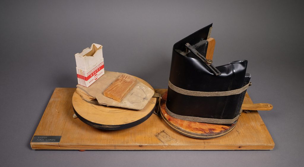
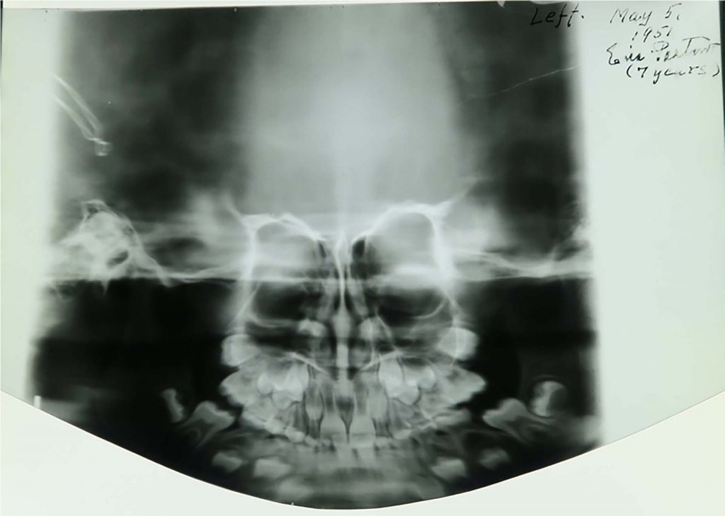
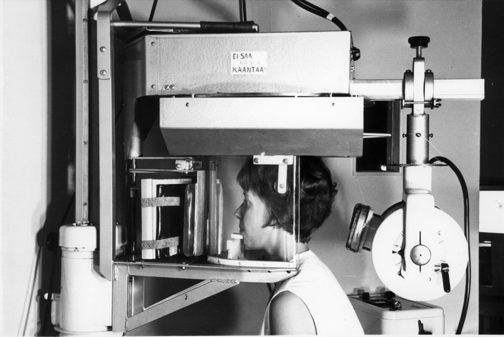
Our object of the month is a trial version of the next stage of the invention, the orthopantomograph, built of parts found in Paatero’s toolbox. This device features two adjacent disks that rotate when the lever is turned. An X-ray film bent in a U shape is placed on the disk on the right, whereas a supporting headrest, made of a small cardboard package for use during a trial session, is set on the left-hand side disk. The prototype was completed in 1957 and used to take the first panoramic X-ray of the skull, providing a right-angled view of each tooth. In the final version, one of which is included in the collections of the Helsinki University Museum, the patient’s chair remained in place, but the film and X-ray machine rotated.
Race to exploit the invention
To develop panoramic radiography, Yrjö V. Paatero had to rely largely on his own funds because he obtained few grants. Of crucial importance was the invitation he received in 1950 based on his articles on the topic to develop his parablograph at the University of Washington in the United States. He worked there for 18 months almost day and night, eager not to waste a moment of the time he had access to all the material resources he needed. Paatero’s work in the United States ended in September 1951. Upon leaving the country, he noticed that all of the documents pertaining to the development of the pantomograph had disappeared from his briefcase.
When he arrived in Finland, he made new designs and drawings from memory, and received funding from the University of Helsinki’s Institute of Dentistry for the construction of the first pantomograph. In the United States, Paatero’s idea failed to advance despite his drawings, designs and material resources because one factor had been missing: a creative problem-solver to address the challenges associated with the idea. The pioneering invention also attracted others keen to exploit it. One collaboration offer that eventually ended in a bitter dispute led to the production of a pantomograph called Rotagraph in England. However, the first orthopantomograph was manufactured in Finland as a result of collaboration between Paatero and Timo Nieminen, an engineer.
A child inventor
Yrjö V. Paatero has described how he invented panoramic radiography on a night in January 1949 when his new-born second child kept the family awake and he decided to use the long hours to invent something. It was by no means the first time he had tried to solve a practical problem or built equipment. Since childhood, Paatero had had a creative streak and enjoyed artistic pursuits, such as drawing, music and poetry, with photography as a special interest. However, as a camera was deemed too complex a device for a child, Paatero had built one himself and also developed photographic plates. He had built other devices, too, such as mechanical toy horses for other children, and later taught his own child to build crystal and transistor radios. Although the panoramic radiograph is his most significant invention, he also developed and applied three-dimensional X-ray imaging.
Longevity based on quality
Yrjö V. Paatero died unexpectedly in February 1963, but in his final years, he had witnessed the importance of his work recognised in many ways. In 1954 and 1955, he received awards at two fairs for inventors, first in Brussels and then in Paris. In 1959 the Finnish president awarded him the title of professor. Moreover, on account of his significant research work, Paatero was invited in 1961 to a professorship in dental radiography established at the University of Turku. He also derived at least a small degree of financial benefit from his invention when the commercial production of the orthopantomograph began in 1961. It is likely that Paatero had a similar attitude to his life’s work as to a piece of petrified wood that he had purchased from the United States and that had holes bored by an ancient worm: “[…] quality work endures even though the worker is lost in the mists of time.”
Helena Hämäläinen, curator
Translation: University of Helsinki Language Services
Sources:
Juhani Wolf, Erkki Tammisalo and Erkki Paatero (2002). Yrjö V. Paatero – Panoraamaröntgenkuvauksen keksijä. In Hippokrates – Suomen Lääketieteen Historian Seuran vuosikirja 2002. Pp. 99–118.
Tulonen, Aimo (1952). Pojan itku sai isän keksimään – Suomalainen röntgenlaite. Päivän Uutiset, 25 June 1955. P. 7.