To fear or not to fear: understanding the world of the Reed Warbler

Hi all!

My name is Sarella Arkkila and I am a second year masters student in Ecology and Evolutionary Biology at the University of Helsinki. I did my bachelor’s in Animal Behaviour at the University of Aberdeen and knew that this is what my passion lies in. Coming to the University of Helsinki, my search for a research group studying animal behaviour began. I found the amazing Information Ecology and Co-evolution research group ( https://www2.helsinki.fi/en/researchgroups/evolution-sociality-behaviour/information-ecology-co-evolution ) led by Prof. Rose Thorogood and knew that this is where I wanted to continue my career within biology. I was honoured to be allowed to join the research group.

My thesis work is on the effect of the landscape of fear on the parenting behaviour of the Eurasian reed warbler. Landscape of fear (LOF) is how animals view their surroundings and the different risks associated within that surrounding. There are several factors that contribute to the LOF, such as the perception of the risk of predators and parasites. LOF in turn influences an animal’s behaviour in different areas of its habitat. For example, in an area where an animal perceives the predation risk to be higher, they may exhibit more vigilance behaviour. These changes in vigilance behaviour can influence foraging efficiency and parental provisioning, as the focus is on spotting and avoiding predation, which can ultimately affect an animal’s fitness. The study of LOF is crucial especially now that environments are changing rapidly, and where new pressures arise and the survival of species within their new or existing habitats are uncertain.

Within the concept of LOF, a few studies have been done on how changes in perceived risk of predation impact indirectly on fitness, but perceived risk of parasitism is yet to be tested. The common cuckoo is a brood parasite, laying its eggs in other species’ nests, one of which is the reed warbler. The reed warbler changes its behavioural defences when it perceives that it is at greater risk of parasitism by cuckoos. This behavioural change can either result from direct observations of seeing a cuckoo or from social information from neighbouring birds attacking a cuckoo in its nest. However it is unknown if this perception of greater risk of parasitism has indirect and long-term effects on behaviour, ultimately affecting the quality of parenting.

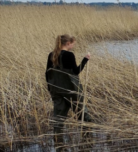
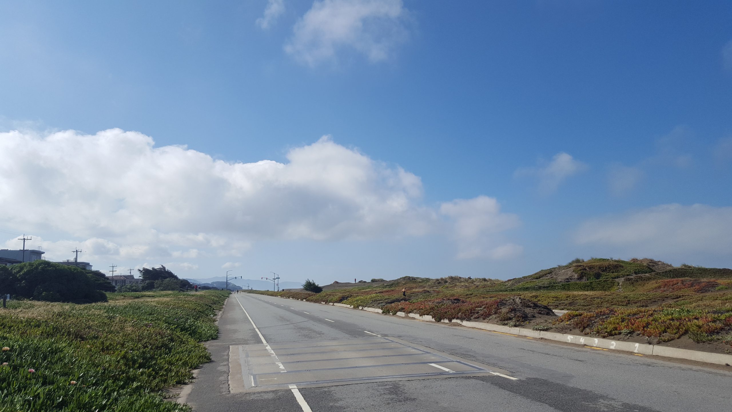
Looking for Reed Warbler nests within the reeds around Helsinki.

To understand the impact of the LOF from parasitism, I conducted fieldwork with the research group in the summer 2021. As part of another experiment, we manipulated the social information (SI) that the birds received about cuckoos by using model nests (previously collected nests), threats (painted 3D models), and alarm call playbacks. I then compared their ‘fear-type’ behaviour both during the presentations and afterwards to birds presented with alarm calls but an innocuous ‘threat’ (a teal), or to ambient teal calls as a control. I collected behavioural observations from video recordings at the nests, which gave me a ‘reed warbler’s perspective’ that could not be gained by observing the birds in person. To evaluate the long-term impact of the LOF, I then recorded parental behaviour 6 days later during incubation, and then again once the chicks had hatched.

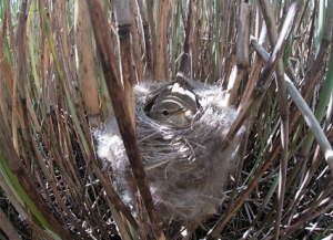
A reed warbler incubating its eggs, a screenshot taken from an incubation video.

For my Master’s thesis, I have been watching a sample of these video recordings to determine how socially-provided information about parasitism risk influences behaviour. Thanks to the HiLIFE traineeship, I can now dive deeper into the videos to find out more about the longer-term effects of fear and publish a scientific article on the findings. I hope to be able to share the study results with you after this fantastic HiLIFE traineeship period. Wish me luck!

HiLIFE TRAINEE ATTENDING THE ASHG21 ANNUAL MEETING

Hi fellow students! My name is Celia Gómez Sánchez, and I am a second-year master’s student in Genetics and Molecular Biosciences. In October 2021 I received the opportunity to attend the American Society of Human Genetics (ASHG) 2021 annual meeting. The conference was held in the US with a hybrid model, so attendees could watch the talks either on-site or online. This enabled people from all over the world to participate ¬–including me. Because there were multiple events happening simultaneously, during the week when the ASHG21 meeting happened I only attended the talks I was most interested in; nevertheless, the conferences were recorded, so they were later accessible to everybody like me that could not watch them live. This was a great feature since I was very keen on most of the topics, and wanted to make the most of this opportunity 😀

As a master’s student, I didn’t present any type of research, I only listened to the conferences. However, this was enough to learn a lot of about various topics, some of which were closely related to my master’s thesis that I’m currently working in. I was especially interested in genetics and neurosciences, and the talks concerning neurodevelopmental diseases were definitely my favorites. We learnt about the use of both cells and mice as models for the different disorders, and how they had explored the genome to find disease-causing variants. For example, one of the groups were able to identify autosomal recessive variants in genes previously unlinked to autism spectrum disorder, resulting in 31% rate of patient diagnosis. Furthermore, the talks explained the use of genomic techniques that I had recently studied (such as single-cell ATAC-seq or single-cell transcriptomics), which provided me with a great opportunity to understand better the application of these techniques with real examples and the results they provide with.

Moreover, some of the meetings specifically focused on craniofacial development, my area of research at the moment. I am studying the cranial neural crest (that gives rise to the craniofacial region), and certain conditions related to defects arising from it, such as pituitary hormone deficiency and maternally inherited gingival fibromatosis. These conditions result in delayed growth and puberty and craniofacial malformations. What is very interesting about the neural crest is that 30% of all congenital malformations in humans are derived from abnormalities in it, since the neural crest precedes the formation of multiple tissues in the embryo. Therefore, its study is key to preventing and treating multiple birth defects, and so I was very excited to learn about state-of-the-art research involving this embryonic structure. As a matter of example, I was glad to hear about TFAP2A, a cranial neural crest marker on which I have been focusing my experiments. I learnt that enhancer mutations that dysregulate this gene were found to cause branchio-oculo-facial syndrome (BOFS), a condition that results in eyes and ears malformations together with characteristic facial features.

In conclusion, being able to attend the ASHG21 meeting was a very powerful experience, that allowed me to learn about multiple topics of interest and get in touch with current methods to research human diseases. I am very happy to have been able to attend these conferences and I really thank HiLIFE for this opportunity, that I wouldn’t have been able to get on my own.

Celia Gómez Sánchez

Attending ESOT-congress as a HiLIFE-trainee


Hi, and welcome to the HiLIFE-trainee blog! My name is Akseli Bonsdorff and I am a fourth-year medical student at the University of Helsinki and also working on my PhD-thesis in pancreas transplantation and general pancreatic surgery. My first scientific article on the role of early plasma amylase levels in predicting pancreas graft-related complications after pancreas transplantation was published in early 2021 and I received an opportunity to present the key findings of the study at the European Society of Transplantation (ESOT) 2021 congress. ESOT is a biannual congress that brings together transplantation surgeons, nephrologists, hepatologists, pathologists, nurses, and students – such as me – and acts as a platform for discussing novel topics and state-of-the-art findings in the field of transplantation.

This year, ESOT was held as a hybrid conference, and attendees had the chance to choose between on-site or online attendance. For approximately ten to twelve hours a day, I sat and listened through the many inspirational talks, keynotes, presentations, and discussions on topics ranging from the definition of brain death to the implementation of artificial intelligence applications in transplant organ allocation systems, and from the effect of Covid19 on the transplant communities to why living donor liver transplantation is not performed extensively in Western countries but cover the majority of liver transplantations in Asian countries. I am still dumbfounded by the exhaustive coverage of different topics, and probably still- after two weeks from the congress – processing all the new knowledge I gained.

On the third day of the congress came the moment of my presentation. Due to the hybrid model, I had to prerecord the bulk of my presentation and my role during the session was to answer questions arising from the audience on-site and online. The two dutch professors chairing my session were not too harsh on me, and one of them even thanked me for an interesting presentation (which he did for every presenter, but needless to say, it felt nice at the moment). Hybrid or not, my debut was something I will remember for a long time.

Without a doubt, ESOT2021 had provided me everything I expected and probably even more. I had the chance to delve deeper into my own field of research (that being pancreas transplantation), but also received tasters into the other subspecialties in transplantation. I am extremely grateful for the financial and scholarly help from my great mentors Adj. Prof. Ville Sallinen and Adj. Prof. Ilkka Helanterä. I also thank HiLIFE for the support received. Making attending an event like this possible for students and aspiring researchers is extremely appreciated.

Akseli Bonsdorff

Fireballs of ischemic stroke

Hi Everyone!

My name is Katariina Järvinen, and I am a second-year master’s student in the Translational Medicine program at the University of Helsinki. I conducted my master’s thesis research during summer 2020. One of the aims of my project was to evaluate the potential of different phagocytosis related genes on enhancement of microglia phagocytosis.

Working as a HiLIFE research trainee in Mikko Airavaara’s research group, I learned many new skills, for example culturing of cells, and gained valuable practical experience from working in a laboratory. In addition, I was able to widen my professional network. Overall, I was impressed with how the whole group works together: if something doesn’t go as planned, there is always help available and multiple people giving ideas on tackling the issue or guidance in proceeding with another suitable method.

Here is a picture of BV2 microglia 1 hour after adding phagocytosis bioparticles. Inside the cells the bioparticles react to pH change and give red fluorescence signal.

Being HiLIFE Research Trainee met all of my expectations despite COVID-19 restrictions. I’m happy that I got to do my traineeship in a research group that is doing research that excites me and that my project has high scientifical value to the members of the group. The results gained during the traineeship will be validated in ischemic stroke in vivo study.

I am deeply grateful to Mikko Airavaara and my supervisor Helike Lõhelaid for their support and guidance during my traineeship and after it. I would also like to thank HiLIFE for supporting me in my early career in medical research.

Here you can read more about neuroprotection and neurorepair group.

Katariina Järvinen

Leiomyomas, bioinformatics, a little bit of lab work and wonderful people

Circos plot

Hi, all of you reading this HiLife trainee blog! I am Vilja and I am a master’s student in genetics and molecular biosciences. At the beginning of this year, I was looking for a master’s thesis position, and I wished to find a project related to cancer or other tumors. I also wanted to further develop my skills on bioinformatics. I was super excited to get a master’s thesis position at Pia Vahteristo’s research group studying gynecological tumor genomics. I was supposed to start with my master’s thesis project in June, but the COVID-19 pandemic changed my plans. In the middle of August, I was happy to finally start with a very interesting project.

In my master’s thesis project, I am analyzing the genomic data of uterine leiomyomas. Uterine leiomyomas are benign smooth muscle tumors of the uterus. The prevalence is up to 70–80% in women. I am focusing on structural variants, such as translocations, inversions, insertions, huge deletions and amplifications. The HMGA2-RAD51B translocation is one of the best characterized structural variants in leiomyomas. This means that part of the chromosome 12 is attached to a part of the chromosome 14, so that HMGA2 receives active regulatory elements of RAD51B. This leads to a much higher expression of HMGA2 in tumor cells compared to normal cells. In addition to this well-known translocation, we have been lucky to come across and characterize some other structural variants in uterine leiomyomas.

Structural variants can be studied in many ways. Sometimes, if you have a hunch of the putative genes mutated and you are lucky enough, you might find something interesting by simply visualizing genomic data on IGV or some other visualizing software showing the alignment of paired-end data. There are also many bioinformatics tools which look for structural variants by utilizing algorithms. These tools, such as Delly, are analyzing discordant reads and split reads of the paired-end data. Also, bioinformatics tools analyzing read coverage can be used to detect copy number variations. Once you have found a structural variant, you may want to validate your finding. This can be done by using PCR and Sanger sequencing. These steps are exactly the ones that I am using for my master’s thesis project.

Circos plots are a nice tool for visualizing these structural variants. In the figure, you can see one of the preliminary Circos plots I have made by using RCircos (R package). Lines show translocations between chromosomes and intrachromosomal inversions, and the circular heatmap indicates copy number variations.

I have learned so many new things during the process, and the project is not even completed yet. For example, I have developed my skills in bioinformatics and learned about genetics and tumorigenesis of leiomyomas. I have also gained a better understanding of how research projects are performed and how technical difficulties can be approached and tackled. I am super lucky that I have had such great supervisors and colleagues, who I really want to thank. Also, I am very grateful for the support I have received from HiLife. It has been extremely valuable.

Vilja Jokinen

DNA, comets and shaky hands

Hi everyone!

My name is Piia Karhu and I am a first-year student in The Master’s Programme in Human Nutrition and Food-Related Behaviour at the University of Helsinki. I received a HiLIFE scholarship for the summer 2020 to work in a research project of my interest. I completed my research internship at the Viikki Molecular Nutrition group. The group studies the molecular mechanisms by which diets, foods and diet-derived compounds mediate their effects on health and prevention of non-communicable diseases. I was very happy to join this group as molecular nutrition is a very fascinating field of science. Here you can read more about Viikki Molecular Nutrition group.

During my summer, I was privileged to take part in the study that examines the impact of dietary habits on faecal water genotoxicity.  In the study, colonial epithelial cells were exposed to feacal water. Feacal water was extracted from stool samples collected during dietary intervention with healthy volunteers and the DNA damage created by the faecal water was examined. Purpose was to study whether there are differences in DNA damage of the cells when exposed to fecal water from volunteers that followed either plant protein diet, animal protein diet or diet that contains half plant protein and half animal protein. We measured the DNA damage using single-cell gel electrophoresis known as Comet assay. It is a commonly used and straightforward method for measuring DNA strand breaks in cells. The term “comet” refers to the leaked-out DNA of the cell as it often resembles a comet. The more intensive the comet tail is relative to the head of the cell, the more the cell is damaged.

microscope image of green cell comets
Here is an example picture of the comets

I had very interesting and mind opening summer job as I have never worked in this kind of project before. I acquired valuable experience working in the lab, especially in the cell lab and learned how to culture and handle cells. Working in a cell lab was a very nice experience even though it took a while before my hands stopped shaking! During my summer I gained a lot of practical experience, which will boost my confidence working as a researcher in the future.

I want to thank my supervisor Anne-Maria Pajari for this opportunity to work in her research group and Hana Slabá for excellent guidance during the summer. Also, I am deeply grateful to HiLIFE for supporting me, this was a very valuable experience!

This summer I didn’t sleep so well  

Sleeping wired

Paavo Teitinen
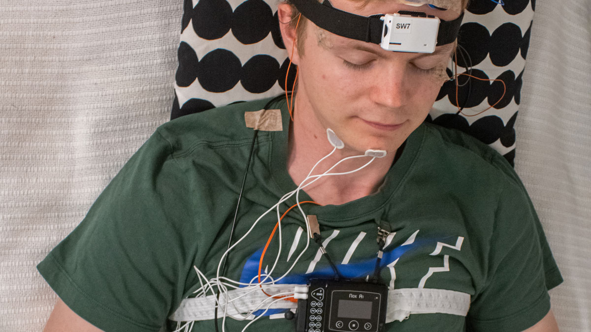
Hi everyone! My name is Paavo and I’m a neuroscience master’s student in University of Helsinki. In this blog text, I will share my experience working as a HiLIFE trainee in summer 2020.

First a little bit of background information. My previous academic background is in exercise physiology. However, even while studying sport sciences I was very interested in neuroscience, especially in the effect of sleep and stress on health. Therefore, I came to continue my studies in Helsinki as there are many excellent research groups here focusing on sleep. That is why I was (and I am) beyond excited, when I got a chance to do my HiLFE traineeship in the Sleep and Health -research group, led by professor Tiina Paunio.

Fun fact: in our first meeting I found out that we both share a background in track and field. I remember Tiina saying: “even though some work may not always be pleasant we can remind ourselves that it is nothing compared to 200 m intervals”. I must agree.

As I had no previous experience in sleep research, my traineeship started with learning polysomnography (PSG) and sleep scoring. PSG is the golden standard of the field, where one attaches a bunch of electrodes to the subject to measure their brain activity, muscle tone, heart rate and breathing. Sleep scoring means determining different macro- and microstructures of sleep from the (PSG) data. I was lucky to have Tuula Tanskanen as my scoring mentor. At first scoring was like being at optician when your glasses are not up to date: Tuula asked what this 30s epoch looks like, I squeezed my eyes looking at the signal, thought a bit and ended up half guessing. Luckily, practice trains your eye to find things from the signal, and I would like to think that the glasses I’m wearing now are at least close to the correct prescription. In addition to learning PSG and sleep scoring, I begun the basics of programming. Even though I’m still at beginner level, programming skills will for sure prove to be useful in further analyses of the sleep data.

charts
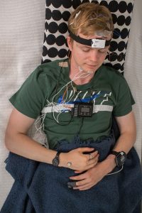
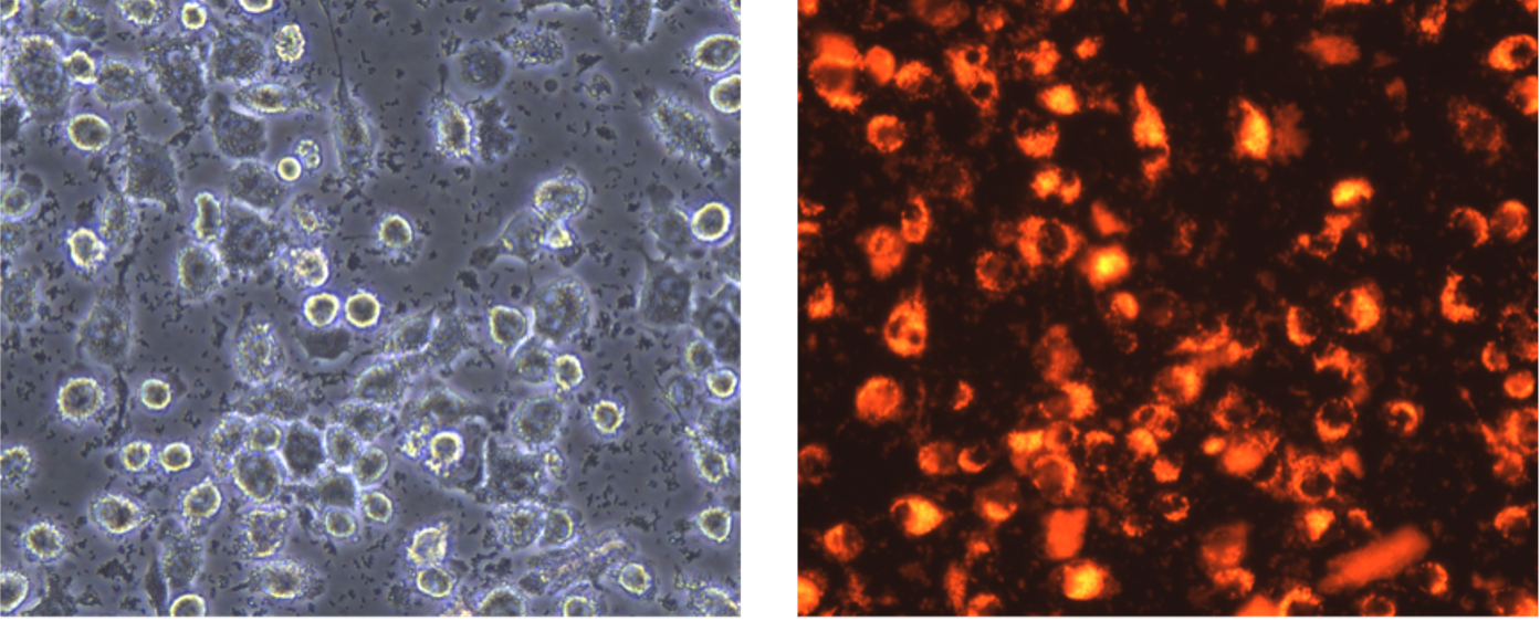
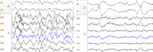
Here is an example of what different sleep stages look like. On the left there is slow wave sleep (N3) with large delta waves on the EEG channels. On the right there is REM sleep with the signature rapid eye movements on the top two EOG channels.

By this time, I’m sure all of you who read the title of the blog are shouting to the screen: “but Paavo why didn’t you sleep well?”. Well I’m glad you asked. You see, in addition to the thesis work I was doing, my job was to test and learn to use some new sleep monitoring devices. These devices will possibly be used in future projects and what better way to learn to use them than to wear them yourself! You can try to guess how many recording devices I’m wearing in the picture and what they are recording. The correct answer will be revealed at the end of this post.

Paavo wired up
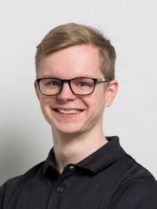
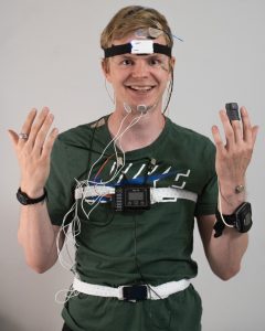
Would you participate in a sleep study if the researcher looked like this?
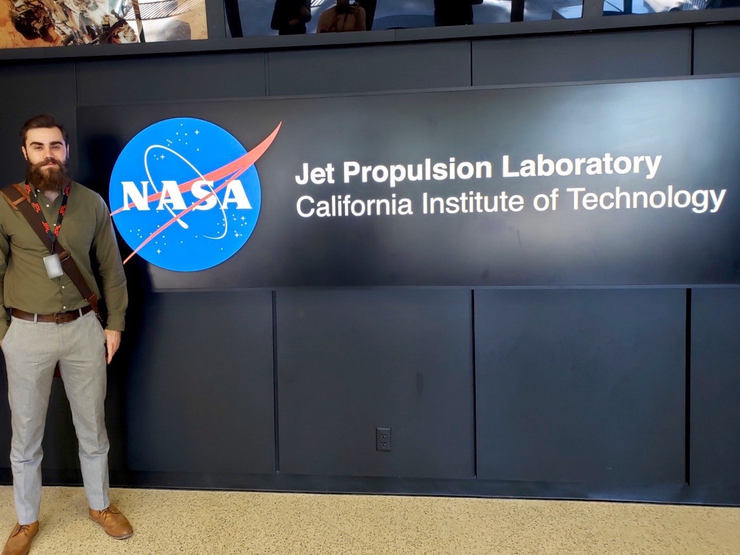
Based on the experience wearing these different devices, I found a direct correlation between the number of cables attached to my head and me being grumpy next morning. The relationship seems to be causal. However, the effect was strongly modulated by the successfulness of the measurement: couple of times I found that nothing was recorded, which was followed by a spike in blood pressure. Testing new equipment always comes with unexpected challenges. Solving these challenges included both reading theory to understand how the devices should work and trying things in practice to solve how they actually work. Couple of times I managed to combine the two: nothing worked, and I had no clue why. Jokes aside, I really enjoyed testing the devices and I could always ask help if needed.

I can honestly say that the HiLIFE traineeship has made a difference for my academic career. Even though I worked most of the time remotely due to covid-19, being able to participate in different (online) meetings and discussing things with my supervisor has opened many opportunities for the future. It is quite likely that I will continue to PhD studies in the same group. A word of advice for future trainees: don’t be afraid to ask and discuss things with members of the research group, you will be surprised where those conversations will lead you. Even the worst-case scenario is rarely worse than running 200 m intervals – and that was one of my favorite track sessions.

Special thanks for my sleep scoring mentor Tuula Tanskanen, for my ask me anything -person Tiina Härkönen and of course for my supervisor and the voice of reason Prof. Tiina Paunio. Thank you HiLIFE for enabling this amazing opportunity.

Paavo wired up and sleeping

  1. For those dying to know, the number of recording devices in the picture was five:

1) The black box on the chest is a polysomnography (PSG) device with several EEG, EOG and EMG electrodes, the white belt and pulse oximeter on the left hand (there are usually even more wring going with nasal airflow sensor and ECG electrodes that are not shown in the picture).

2) The white device on the forehead is an EEG headband, which measures brain activity, movement and temperature.

3) The watch-looking device on the right hand is an actigraphy monitor, which is an accelerometer measuring movement throughout the day.

4) The ring on the right-hand index finger measures heart rate, movement and temperature.

5) The small button on right hand (in the first picture on left hand – the deceit is revealed, the pictures were staged) is a tiny thermometer measuring changes in temperature.

Six months later. A brief reflection.

Arriving at the Baraban Epilepsy Research Laboratory typically involves either climbing eight flights of stairs or waiting an unsettlingly long time while the elevator, long overdue for repairs, shudders its way to the desired floor. My lethargy usually precludes expediency, and I regularly find myself squished in at the back of the enclosed container. On the plus side, the elevator talk is often entertaining; grumblings over the embarrassment that is the elevator, this effectively lightens the figurative load.

Continue reading “Six months later. A brief reflection.”

Turtles, Herpes and Sunshine at Eastern Florida

If someone had asked me five years ago, I probably couldn’t have guessed that I would be spending my sixth year of veterinary studies thousands of miles away from home, sitting inside a lab with an air conditioning as enthusiastic as October winds in Helsinki. Yet there I was, soaked in the familiar smell of Clorox wipes, whirring tiny tissue bits through various machines in the slow spurring excitement of soon finding out something, even small, that no one else ever has before.

Continue reading “Turtles, Herpes and Sunshine at Eastern Florida”